Download
1 / 24
240 likes | 589 Views
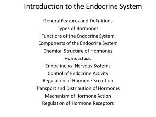
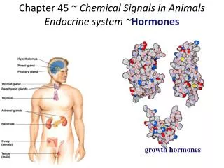
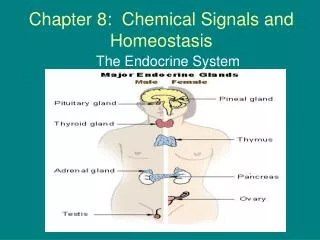
Chapter 45 Chemical Signals in Animals; Hormones and the Endocrine System. Michael Kosofsky and Emily Pipilas. Comparison. Endocrine System Hormones travel in blood to target cells Controls tissues Regulates metabolism Feedback in seconds to months. Nervous System Electrical signals

E N D
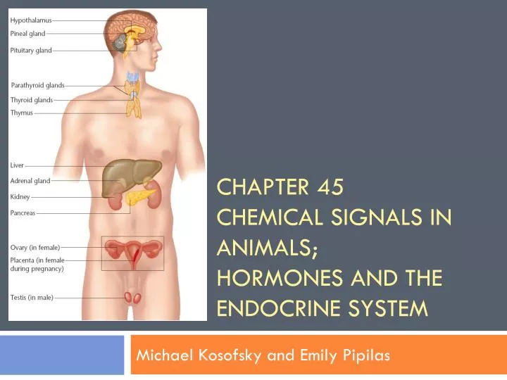
Chapter 45Chemical Signals in Animals;Hormones and the Endocrine System Michael Kosofsky and Emily Pipilas
Comparison Endocrine System • Hormones travel in blood to target cells • Controls tissues • Regulates metabolism • Feedback in seconds to months Nervous System • Electrical signals • Release neurotransmitters to synapse • Controls muscles and glands • Quick feedback
Feedback Mechanisms • Negative Feedback • Primary mechanism of homeostasis • Change in physiological variable triggers response that counteracts initial ‘away from normal’ fluctuation • Positive Feedback • Physiological control mechanism • Change in variable triggers mechanisms to amplify that change
Steroid Model vs. Protein Model Steroid Model Protein Model • Diffuse through cell membrane • Receptors in nucleus • Response has slow onset • Long-lasting • Typically derived from cholesterol • Examples: Androgens, Estrogens, Progesterone • Don’t diffuse through cell membrane • Receptors on surface of cell • Response has quick onset • Short-lasting • Typically derived from amino acids • Examples: GH, Insulin, Glucagon
Paracrine Signaling: local signaling between nearby cells within organ (don’t travel in blood) • Endothelial cells of blood vessels release nitric oxide to help relax the smooth muscle in blood vessels, thus dilating blood vessels • Smooth muscle cells also produce prostoglandins, which aid in smooth muscle contraction • Examples: uterine wall contractions
3 Stages of Cell Signaling • Reception: occurs when signal molecule binds to specific receptor protein • Receptor proteins are built into the plasma membrane of the target cell or located inside the target cell • Target cell: Cell that responds to a regulatory signal such as a hormone • Signal Transduction: events within the target cell that are triggered from the binding of a signal molecule to a receptor protein • Response: change in the cell’s behavior
Ligand • Ligand: molecule that binds specifically to the receptor site of another molecule • 3 Models for Ligand/Receptor Interactions • G-Protein Linked Receptors • Tyrosine-Kinase Receptors • Ion-Channel Receptors • Intracellular Receptors
Ligand-Gated Channel • Protein pores in the plasma membrane that open or close in response to a chemical signal, allowing the blocking or flow of specific ions such as sodium or calcium ions. • Bind to signal molecule as a ligand at a specific sight on their extracellular side. • The shape change produced in the channel protein immediately leads to a flow of particular ions along their concentration gradient through the channel.
Steroid Hormones • Derived from cholesterol • Lipid soluble (able to diffuse through cell membrane) • Sex hormones • Progesterone • Estrogen • Testosterone • Adrenal Cortex Hormones • Actions • Hormone crosses membrane • Hormone combines with receptor in nucleus • Attaches to section of DNA • Transcription (synthesis of mRNA) activated • mRNA enters cytoplasm to ribosome to create protein
Second Messenger • Chemicals that transfer and amplify the signal from the receptor to proteins • Cause a specific response
Calcium Ions and InositolTriphosphate IP3 in Signaling (Secondary Messengers) • Signal molecule (first messenger) binds to G-protein-linked receptor, leads to activation of phospholipase C • Phospholipase C cleaves a plasma membrane phospholipid called PIP2 into DAG and IP3 • DAG functions as a second messenger in other pathways • IP3 diffuses through cytosol, binds to a ligand-gated calcium channel in the ER membrane, causing it to open. • Calcium ions flow out of ER (down concentration gradient) raising calcium ion level in cytosol • Calcium ions activate the next protein in more signaling pathways, often acting via calmodulin, a calcium ion-binding protein
Cellular Response to Cell-Signaling Pathway • Response of liver cells to signaling by the hormone epinephrine • Helps regulate cellular energy metabolism • Activates the enzyme that catalyzes the breakdown of glycogen
Duct vs. Ductless Glands Duct Glands Ductless Glands • Exocrine • Secrete their products into ducts • Lead directly to external environment • Examples: sweat glands, salivary glands, mammary glands • Endocrine • Do not possess any ducts by which secretions are discharged • Secrete hormones directly into bloodstream • Examples: thyroid, parathyroid, thymus, pituitary body, pineal body
Anterior Pituitary Hormones • Growth Hormone (GF) • Endorphins • Prolactin (PRL) • Follicle-Stimulating Hormone (FSH) • Luteinizing Hormone (LH) • Thyroid Stimulating Hormone (TSH) • Adrenocorticotropic Hormone (ACTH) • Melanocyte-Stimulating Hormone (MSH)
Hypothalamus-region of lower brain that recieves information from nerves throughout the body and from other parts of the brain then initiates endocrine signals appropriate to environmental conditions -secretes hormoones of the posterior pituitary and releasing factors, which regulate the anterior pituitary • Neurosecretory cells • Influence the pituitary gland which integrate endocrine and neural function • Posterior Pituitary: extension of hypothalamus that stores and release two hormones (oxytocin and antidiuretic hormone) • Anterior Pituitary: Releases and inhibits hormones conveyed by portal vessels in the hypthalamus. Hormones include: TSH, FSH, LH, GH, PRL, ACTH, MSH, endorphins
Antidiuretic Hormone • Release of ADH from the anterior pituitary is inhibited by drinking alcochol • How would this affect urination?
Thyroid Gland • Releases Calcitonin when blood calcium levels are too high, lowering calcium levels in the blood • Osteoblasts build bone • Produces T3 and T4 (iodine-containing hormones) • Stimulate metabolism • Influence development and maturation Parathyroid Gland • Secretes Parathyroid Hormone (PTH) • Raises calcium levels in blood plasma when they are below normal • Works with calcitonin to maintain blood calcium homeostasis • Stimulates kidneys to activate Vitamin D, which causes intestines to increase uptake of calcium from food
Iodine Deficiency in Diet • Results in goiter • Enlargement of thyroid • In the absence of sufficient iodine, thyroid gland cannot synthesize adequate amounts of T3 and T4 • Leads to continuous secretions of TSH, which then leads to an enlarged thyroid
Thymus Gland • Active in establishing the immune system • Secretes thymosins • Promotes the development of white blood cells (lymphocytes) • Stimulate T cells (immunity)
Pancreatic Hormones & the Regulation of Blood Sugar (Negative Feedback) Insulin Glucagon • Released when blood sugar levels are high • Stimulates cells to • Perform cellular respiration • Turn glucose into glycogen (where it is stored) • Turn excess glucose into fat • Take in amino acids for protein synthesis • Convert fatty acids into lipids • Released when blood sugar is low • Liver- change glycogen into glucos • Adipose (fat) tissue- breakdown of fats and proteins to form glucose
Stimulation of the Adrenal Gland • Hypothalamus • Neural Signals • Sympathetic Impulses • Adrenal Medulla (medial) • Epinephrine and Norepinephrine Released • Short term “fight or flight” or alarm stage • Hypothalamus • CHR Released • Anterior Pituitary • Releases ACTH • Adrenal Cortex (lateral) • Releases Cortisol • Long term Adjustment or Resistance Stage
Epinephrine • Causes short term “fight or flight” or alarm stage • Blood glucose increases • Blood glycerol and fatty acids increase • Heart rate increases • Blood pressure rises • Breathing rate increases • Air passages dilate • Pupils dilate • Blood flow redistributes, leading to increased alertness and decreased digestive and kidney activity
Gonadotropic Hormones FSH LH • Stimulates gamete formation • Female: follicle, egg-maturation in ovary, release of estrogen • Male: sperm production in tubes of testes, release of testosterone • Negative Feedback: Release of Gonadotropin Releasing Hormone (GnRH) from the hypothalamus-decreased when levels of progesterone and testosterone rise • Stimulates ovulation in the ovary (female) • Ovulation causes release of progesterone • Causes production of testosterone in testes (male) • Negative Feedback: Hypothalamus decreases its release of GnRH when levels of progesterone and testosterone rise
Secondary Sex Traits Secreted by Ovaries (Female) Secreted by Testes (Male) • Estrogen • Support egg maturation • Controls period • Testosterone • Sperm production • Protein synthesis in muscle