Download
1 / 14
140 likes | 163 Views
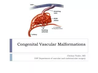
Learn about arteriovenous malformations, venous angioma, cavernoma, capillary teleangiectasia, and more non-neoplastic brain vascular malformations. Explore their pathophysiology, clinical presentation, and imaging studies in this informative guide.

E N D
Cristina Caterina Aldea Scientific Coordinator: Professor Ioan Ştefan Florian MD, PhD “Iuliu Haţieganu”University of Medicine and Pharmacy Cluj-Napoca Vascular malformations. General considerations
Non-neoplastic Vascular Malformations of the Brain • Arteriovenous Malformations (AVMs) • Venous Angioma • Cavernoma • Capillary Teleangiectasia • Direct Fistula ( McCormick-1966 )
Clinical presentation 1. Hemorrhage - most common - 82% significant IP component - SAH, IVH 2. Seizures 3. Mass effect (increased ICP ) 4. Progressive Neurologic Deficit 7% of patients with AVMs present associated aneurysms
Imaging studies • CT scan • CT angiography • MRI / angio - MRI • Cerebral angiography
Cavernomas = cavernous angioma, cavernous hemangioma
Clinical presentation • Seizures • Progressive Neurological Deficit • Hemorrhage ( usually IP ) > 50 % asymptomatic
Imaging Studies • CT • MRI - hemosiderine ring( “salt and pepper” appearance ) • Angiography – “angiographically occult”