Download
1 / 35
441 likes | 1.17k Views
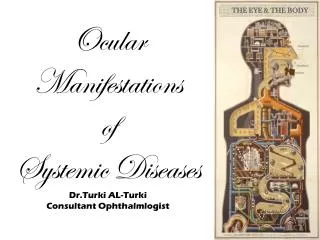
Ocular Manifestations of Systemic Diseases. Dalman. Dysthyroid Orbitopathy. autoimmune disorder usually associated with Graves' disease 10-25% euthyroid extra-ocular muscles are the target of the autoimmune attack restrictive ophthalmoplegia and proptosis Cardinal Signs

E N D
DysthyroidOrbitopathy • autoimmune disorder usually associated with Graves' disease • 10-25% euthyroid • extra-ocular muscles are the target of the autoimmune attack restrictive ophthalmoplegia and proptosis • Cardinal Signs • upper eyelid retraction and lag, conjunctival injection and chemosis, and periorbital edema.
DysthyroidOrbitopathy • Pathophysiology • antibody-mediated reaction against the TSH receptor with orbital fibroblast modulation of T-cell lymphocytes
DysthyroidOrbitopathy • Pathophysiology T- cells inflammation Thyroid cells Hyperosmotic shift Orbital fibroblast EOM edema cytokines mucopolysaccharides
DysthyroidOrbitopathy • Pathophysiology Preadipocyte fibroblasts adipocytes Inc. orbital volume Inc. fat proptosis lagophthalmos edema Tissue damage and fibrosis EO motility restriction
DysthyroidOrbitopathy • Management • self-limited (over 1 year) • No immediate cure available
DysthyroidOrbitopathy • Management • Orbital radiation • moderate-to-severe inflammatory symptoms, diplopia, and visual loss in patients with TAO • Optic nerve compression • High-dose steroids (proceed to surgery if unresponsive) • Surgical • Orbital decompression • Strabismus surgery • Lid lengthening • Blepharoplasty
Occular Changes in Hypertension • Damage to the retina caused by high blood pressure • 3 manifestations • Hypertensive retinopathy • Hypertensive optic neuropathy • Hypertensive choroidopathy
Occular Changes in Hypertension • Pathophysiology • Retinal microvasculature Inc BP Hyperoxic & hypercapneic stress Bifurcation angles and retinal arteriolar diameters show Dec vascular reactivity Disadvantageous branching geometry in retinal vasculature
Occular Changes in Hypertension • Pathophysiology • Dynamics of ocular blood flow Hypertensive arterial changes Inc resistance to optic nerve head blood flow Inc BP Breakdown of autoregulation
Occular Changes in Hypertension • Pathophysiology • Different manifestations because • Acute HTN disrupts blood-retinal barriers • Retinal and optic nerve head vascular beds have autoregulation (choroidal has none) • Choroidal vessels has no blood-ocular barrier • Retinal vessels (no autonomic nerve supply) • Choroidal vessels (richly supplied by both sympathetic and parasympathetic nerves)
Hypertensive Retinopathy • Represents target-organ damage
Hypertensive Retinopathy • Clinical features • Vasoconstriction • Fundus • focal and generalised arteriolar narrowing, microaneurysms, intraretinal hemorrhages, cotton-wool spots, hard exudates, optic disc swelling • 2o to arteriolosclerosis arteriovenous nipping • Flame-shaped hemorrhages (abnormal vascular permeability) • Macular star (lipid deposition around the fovea) • Disc swelling (minimal microvascular change)
Hypertensive Retinopathy • Clinical features • Vasoconstriction • Fundus • Untreated hypertension hemorrhagic detachment of retina and vitreous hemorrhage
Hypertensive Retinopathy • Clinical features • Vasoconstriction • Fundus • Secondary arteriosclerosis • Bonnet’s sign - banking of the venule distal to the crossing • Gunn’s sign - nipping of the blood column • Salus’ sign - displacement of the venule at right angles to the arteriole
Hypertensive Retinopathy • Gunn’s sign and Bonnet’s sign
Hypertensive Retinopathy • Focal arterial narrowing of the retina
Hypertensive Optic Neuropathy • Papilloedema or bilateral disc swelling • Grade IV hypertensive retinopathy • Poor prognostic sign • Other causes like space-occupying lesions and benign intracranial HTN should be excluded • Theories on the pathophysiology • Ischemia and raised ICP as a part of hypertensive retinopathy/enchephalopathy
Hypertensive Optic Neuropathy • Usually resolve following control of BP, but some might develop disc pallor • Longstanding uncontrolled HTN retinal nerve fiber loss
Hypertensive Choroidopathy • Less well recognized than retinopathy • Commonly described features: • Choroidal vascular sclerosis • Elschnig spots – focal areas of degenerative retinal pigment epithelium • Siegrist’s streaks – linear pigment epithelial changes • poor prognosis
Hypertensive Choroidopathy • Elschnig spots
Management • Control hypertension • Grade I and II • Non-urgent referral • Grade III • More urgent referral to the GP • Grade IV • Patient is in medical crisis. Patient needs immediate referral to a hospital eye casualty department