Download
1 / 80
1.2k likes | 2.46k Views
Pathophysiology of vascular tone. Arterial hypertension. Prof. Olha V. Denefil.

E N D
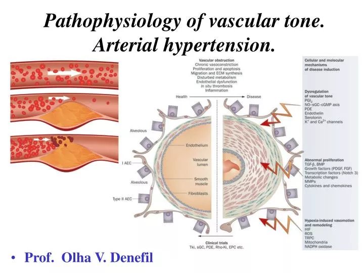
Pathophysiology of vascular tone. Arterial hypertension. • Prof. Olha V. Denefil
Increased blood pressure is found in 15-30 % of the adult population in the world. There are significant differences in this indicator over the world: from 6 % - in Africa to 30-35 % - in the Scandinavian countries, in the USA is 23-31 %, in Ukraine – nearly 40 %.
Arterial hypertension is a major risk factor of many pathological conditions and diseases of the cardiovascular system: atherosclerosis left ventricular hypertrophy and heart insufficiency, ischemic heart disease (myocardial infarction) cerebrovascular disease (ischemic and hemorrhagic brain stroke) renal insufficiency
„RULE of HALF” Thus, only about 25 % of patients taking medications to lower blood pressure About 50% of people do not know about an increase of their blood pressure!!! Of those who know 50% of untreated!!! Effective antihypertensive therapy have only 12-13 % of patients
REGULATORY SYSTEM, THAT PROVIDE STABILITY OF BLOOD PRESSURE Haemodynamic systems Systems of control Stable Arterial Pressure
Systems of AP control SYSTEM of BRIEF ACTION SYSTEM of LONG TERM ACTION Baroreceptors and chemoreceptors of aortic arch and sinocarotid zones renin - angiotensin II - arterioles angiotensin II aldosterone
Classification Arterial hypertension AP above 139/89 mm Hg Arterial hypotension Primary (essential) Secondary (symptomatically) AP less than 100/60 mm Hg Acute (collapse) Chronic (hypotonic disease, symptomatically)
Classification Primary AH (essential, hypertonic disease) 80 % of all increase of AP Secondary AH (that is happened in 5 -10 % cases). It’s a symptom of some disease course
Etiology of primary AH Reason is unknown (AH is polyetiological disease). AH arises on the ground of genetically peculiaritiesof metabolism. That is possible to have genetically defect of the systems, which control relaxation of the smooth muscle cells of the arterioles. It is possible to: 1. Hereditary defect of Ca-ATPase (myocyte relaxation of arterioles involves the movement of Ca in the sarcoplasmic reticulum against the concentration gradient) 2. Genetically caused sodium retention in the body 3. The variability of the gene that controls the synthesis of receptors for angiotensin 2 4. Genetically caused by increased activity of ACE 5. Lack of endothelial nitric oxide synthesis
Theories of primary hypertension • 1. Recognized the leading role of the nervous system • Essence: disorders of the nervous regulation of vascular tone • against failure mechanisms of hormonal regulation of arteriolar tone carried over earlier kidney disease, age-related changes in blood vessels, endocrine disorders during menopause • 2. Recognized the leading role of the kidneys • Essence: imbalance pressor and depressor functions • Increased vascular tone occurs on the background of exhaustion depressor of kidneys
Contributing factors Age-related changes in blood pressure High salt intake Stress Family history Obesity (hyperinsulinemia) Excess alcohol consumption (mechanism in unclear) Hyperinsulinemia: causes high activity sympathetic link of ANS and its effect on cardiac output, peripheral vascular resistance and renal sodium retention; stimulates sodium and calcium transport across the cell membrane of vascular smooth muscle, thereby sensitizing blood vessels to vasopressor stimuli Race (for example: AH isn’t only more prevalent in African Americans than whites, it is also more severe). Possible explanation: due to evolutionary adaptation to the severe environment (western Africa and Western hemisphere) in condition of salt and water deprivation survival is possible due to retention of sodium and water in organism. That leads to conserve sodium. There is little information about other racial groups
Pathogenesis 1. Increased blood volume NaCl (use of more than 5 g per day) - mountain population of Japan, the Ukrainian Carpathian and Crimean often suffer from hypertension disease due to the use of water that contains a lot of NaCl Causes Reduced of Na+ excretion by the kidneys (kidney disease) Genetically caused decrease Na excretion by the kidneys
EtiologysecondaryАH • Renal • (resulted from kidney pathology) Glomerulonephritis Kidneydamage at collagenosis Uri stone disease Kidneyamiloidosis Kidneytumor Glomerulosclerosis because diabetes mellitus Nephropathy of the pregnant Hereditary defect of renal vessels Renal vessels atherosclerosis, embolism or thrombosis
EtiologysecondaryАH 2. Renoprive (arises afterkidney remove) 3. Angiogene (is caused by vessels pathology) Aorta damage Arteries carotids damage
EtiologysecondaryАH 4. Endocrinopathy (develops in the result of endocrine glands pathology) Acromegaly (Somatotropin over production by the pituitary gland anterior part) Hyperaldosteronism (aldosteronover excretion by suprarenal glands) Cushing's disease (Adrenocorticotropin over productionby the pituitary gland anterior part) Menopause (age-depended decrease of female gonads activity – estrogens excretion decrease) Possible mechanism – deficit of NO synthesis by endotheliocytes
EtiologysecondaryАH 5. Neurogene (is accompanying to nerves system pathology) Encephalitis Brain tumor Braintrauma Brainischemia Brainhemorrhage
EtiologysecondaryАH 6. Cardiac Heart defect Heart failure 7. Drug-induced Drugs, which cause vessels spasm (influent on kidney), hormonal contraceptives
Pathogenesis Increase of circulative blood volume (CBV) Emotional excitement (SNS activation) Cardiac output (CО) increase Peripheral vessels resistance increase Kidney functions violation
Pathogenesis Increase of circulative blood volume (CBV) NaCl (intakemore 5 g/day) Reasons Decrease Naexcretion by kidney (kidneydiseases)
Pathogenesis 1. CBV increase Na accumulation in vessels smooth muscle walland increase of its osmotic pressure Na+retention in blood Blood osmotic pressure increase Vessels wall edema Vessels smooth musclesensitivity to vasoconstrictive influences increase (noradrenalin, adrenalin, endotheline, angiotensin) Hypervolemia Vessels narrowing Cardiac output increase Vessels spasm Peripheral vessels resistance increase AP elevation Formula: АP = CO · PR
Pathogenesis 2. Cardiac output increase (CO) Circulative blood volume increase (CBV) Reasons Emotional stress physical (overload) stress Hyperthyreosis
Pathogenesis 2. Cardiac output increase SAS activation Adrenalin excretion Increase of cardiac contractility force Increase of heart beats Increase of cardiac output AP elevation Formula: АP = CO · PR
Pathogenesis 3. SAS activation SAS activation Suprarenal glands activation Interaction adrenalin and alpha-adrenoreceptors Venues and veins smooth muscles spasm Production of catecholamine Noradrenalin Аdrenalin Arterioles smooth muscles spasm Increase of circulative blood in big blood circle adrenoreceptors of heart alpha-adrenoreceptors of vessels Arterioles vasoconstriction Increase of CBV Arterioles vasoconstriction PR increase CO increase CO increase AP increase Formula: АP = CO · PR
Pathogenesis 4. Kidney functions violation Long time spasm of kidney’s arteries • Angiotensin 2 effects • Smooth muscles contraction in the vessels • Stimulation of the vasoactive center in brain • Noradrenalin excretion increase • Adrenalin excretion increase from suprarenal glands • Aldosteron excretion increase from suprarenal glands(Na retention due to kidney) • Activation of Na and water reabsorption in the kidney without aldosterone AP decrease in renal capillaries Activation of JGA Renin excretion Angiotensin 2 synthesis AP increase
Forms of hypertensive disease Norm concentration of rennin in blood (55-60 %) Decrease concentration of rennin in blood (25-30%) Increase concentration of rennin in blood (10-20%)
Depressive function of kidney – synthesis of the substances for AP reduce dilates renal arteries, reduces renin synthesis and reduces Na reabsorbing in kidney PG Е 2 ! ! ! Exhaustion of kidney depressive function leads to arterial hypertension stabilization Phospholipids Renin Inhibitor Angiotensinase Phosphatydilcholin alkali ethers
Arterial hypertension after-effects 1stperiod functional violations (heart hypertrophy) • 2dperiod • Pathological changesin arteriesand arterioles (dystrophy): • Arterioles sclerosis • Arteriole’swall infiltration by plasma (leads to dystrophy) • Arterioles necrosis (hypertonic crisis arisesin clinic) • Vein’s wall thickening
Arterial hypertension after-effects 3dperiod Secondary changes in organs and systems CNS – brain hypoxia – neurons destruction – apoplexy (because vessels destruction and rupture leads to brain hemorrhagesand brain destruction) Kidney (nephrosclerosisandchronic kidneyinsufficiency) • Organs of vision • retinopathy (retina’svessels injury) • hemorrhages and separation (exfoliation) of retina, that leads to blindness Heart Decompensate heart failure Endocrine system Glands atrophy and sclerosis
Pathogenetic principles of treatment 1. Decrease of consumption and increased excretion of fluid and Na+ - decrease of CBV and vascular sensitivity to pressor effects • 2. Decreaseof emotional and physical stress - decrease CNS activity (including sympathoadrenal activity) • 3. Block of adrenoreceptors - reduce the effects of catecholamine in the heart, i.e. CO • 4. Block of adrenoreceptors - reduce the effects of catecholamines in the arterioles, i.e. reducing vasomotor arteriolar tone • 5. Block of ACE - reduce the formation of angiotensin 2 - decrease basal arteriolar tone • 6. Decreasethe effects of angiotensin 2 - blocking receptors for angiotensin 2 - decrease basal arteriolar tone • 7. Decreaseadmission of Ca in myocytes of arterioles - decrease basal arteriolar tone • 8. Increase of depressor kidney function - decrease basal arteriolar tone • 9. Increase of vasodilator function of blood vessels involving nitric oxide - reducing basal arteriolar tone
Plasma lipoproteins are produced and secreted by the liver parenchymal cells and epithelial cells of the small intestine.
The outer membrane of lipoprotein is hydrophilic and inner core is hydrophobic. Lipoproteins are soluble in water, it is a transport form of lipids in the blood. General structure of lipoprotein. There is a lipid drop inside (nucleus), which contains triglycerides (TG) and cholesterol esters (ACh). Membrane covers the nucleus and consists of protein (apoprotein, or apo-), phospholipids (PhL) and non-ester cholesterol (NACh).
In plasma of healthy people is • 4-8 g/l - total lipids • 0.8-1.5 g/l - VLD • 3,2-4,5 g/l - LDL • 2,7-4,3 g / l - HDL • 3,9-6,5 mmol/l -general chylomicrons
Kinds of the lipoproteins Indexes Chylomicron VLDL, pre--LP LDL, -LP HDL, -LP Diameter, nm 500-1000 25-75 19-24 6-12 Chemical structure (%): Cholesterol 0,5- 3 15-17 35-48 20-37 %Cholesterol esters 46 57 66-70 78 Phospholipids 3-9 13-20 11-30 24-40 Triglycerides 80-95 50-70 5-10 3-5 Protein 1-2 5-12 14-25 45-55 Apoproteins A, B, C, E B, C B, C, E A, C, D, E
The value of cholesterol • 1. Necessary for maintaining of cell shape • 2. Together with PL and proteins provides selective permeability of cells to different substances • 3. Source of sex and steroid hormones • 4. Source of bile acids • 5. Necessary for growth of the organism and cell division
Balance of cholesterol • One day in the human body • 450 mg of cholesterol oxidized to bile acids • 450 mg of cholesterol excreted with faeces • 100 mg of cholesterol excreted with dermal fat • 300 mg of cholesterol derived from food • 700 mg of cholesterol is synthesized from acetyl-CoA in the cells of various organs, the highest in the liver and small intestine • In adult is about 140 grams of cholesterol (93% is in the cells, 7% is transported in the form of LP mainly LDL in plasma).
Skin fibroblast Lymphocyte Macrophage Smooth muscles cell of artery Role of LP in Cholesterol transport inside the cell.That is due to receptor-mediated mechanism.It was discovered byAmericanscientists M.Brown and J.Goldsteinin 1973-1975(Nobel Prize in 1985) Аpо-В-receptor Аpо-Е-receptor (receptor connects the LDL, depends on Cholesterol needs of the cell)