Download
1 / 47
500 likes | 802 Views
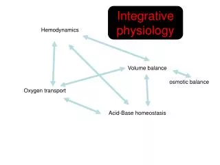
Assessment of perioperative hemodynamics. S.Zahra Ojaghi Haghighi .MD, FACC. Hemodynamic Data Obtainable with 2-D Doppler Echocardiography. Volumetric measurement Pressure gradients Valve area Intracardiac and pulmonary artery pressures Ventricular dp/dt.

E N D
Assessment of perioperative hemodynamics S.ZahraOjaghiHaghighi .MD, FACC
Hemodynamic Data Obtainable with 2-D Doppler Echocardiography • Volumetric measurement • Pressure gradients • Valve area • Intracardiac and pulmonary artery pressures • Ventricular dp/dt
Assessment for Accurate Doppler Stroke Volume Calculations • Blood flow is laminar with a spatially flat flow velocity profile. • Measurements of the velocity-time integral and cross-sectional area(i.e,diameter) are made at the same anatomic location. • The velocity-time integral measurement represents the average velocity-time integral(several measurements shouId be averaged for a patients in normal sinus rhythm,whereas 8 to 10 should be averaged for a patient in atrial fibrillation. • The velocity- time integral is measured with doppler beam parallel to blood flow
Calculation of Cardiac Output • Usually measured at LVOT or aortic valve in the absence of AI. • Correlate well with thermodilution. • Doppler TEE has been used for continuous measurement of CO • CO=SV*HR • SV=CSA*VTI • CSA=0.785*D(LVOT)*D(LVOT)
Method • LVOT(probe in the stomach,turned leftward,in the flexed position): • 1-Parallel pulse Doppler of LVOT • 2-Diameter obtained from either • the stomach or the midesophageal 120- • degree view • RVOT by pulse wave • Pulmonary artery by pulse wave • Aortic valve by continuous wave(cross-setional area of the aortic valve during mid systole) • Mitral valve(CSA=0.785*D1*D2)
LVOT Stroke Volume Calculation Pulse wave SV in LVOT proximal to the AV(approximately 1cm) Determination of the LVOT VTI: Transgastric long axis view or deep transgastric long axis view Diameter of LVOT: midesophageal long-axis view of aortic valve(approximately 1 cm) SV=CSA*LVOT VTI
Practical consideration • For intraoperative TEE ,LVOT is the most reproducible. • Accuracy is improved by assessing multiple Doppler flow profiles,typically 3-5 for a regular rhythm and 10 for an irregular rhythm.
Pulmonary –Systemic Flow Ratio(QP/QS) • Indicates: Magnitude of a shunt(ASD,VSD, PDA) Timing of surgery
Method • Systemic SV(at LVOT or AV) • Pulmonic SV(at PA or RVOT) • Qp/Qs=(PA SV * HR) / (LVOT SV * HR) • QP/Qs=PA SV / LVOT SV
Doppler Measurement of Regurgitant Volume and Fraction • Volumetric Method • Proximal Convergence Method
Assessment of Mitral Regurgitation by Volumetric Method • RVmv=SV mvi – SV lvot • RVmv(%)=(RVmv/SVmvi)*100% • Perform infrequently during TEE due to time and possible error in SVmvi
Assessment of Mitral Regurgitation by Proximal Convergence Method(PISA) • PISA flow=MR flow • 2* 3.14 * r2 * PISA velocity=EROA * MRv • 6.28 * r2 * Aliasing velocity=EROA * MRv • EROA=PISA flow rate/Regurgitant velocity • EROA=(6.28 * r2 *Aliasing velocity)/MRv • RV=EROA * VTI(reg jet)= (6.28 * r2 *Aliasing velocity * VTIreg jet)/MRv
Method • Color flow imaging of PISA from MR • PISA radius • Aliasing velocity • Continuous Dopplerof MR jet to measure peak velocity and VTI of MR
Simplified proximal convergence method • Based on the assumption: • MR velocity =5 m/s • Aliasing velocity is set at 40 cm/s • EROA=r2/2
Doppler Measurments of Pressure Gradients • Bernoulli euation: • dP= 4(V2-V1)2 • dP=4(V2)2
Doppler Determination of Valve Area • Continuity equation: • SV1=SV2 • CSA1 * VTI1=CSA2 * VTI2 • CSA2=CSA1 * (VTI1/VTI2)
Doppler Determination of Valve Area • Flow Convergence Method: • CSA=PISA flow/peak velocity • CSA= (6.28 * r2 * Aliasing velocity/VMS)
Pressure Half-time • Defined as the time required for the peak pressure gradient to decline by 50% • MVA(cm2)=220/PHT • PHT(msec)=0.29 * DT(msec) • AI severity and acute AI(<250msec)
Intracardiac Pressure • RVSP=4(VTR)2+RAP • RVSP=SBP - 4(Vvsd)2 • MPAP=4(Vearly PI)2 + RAP • PADP=4(V late PI)2 +RAP • LAP=SBP – 4(VMR)2 • LVEDP=DBP – 4(Vend AI)2
PVR • PVR=TRV / VTI RVOT* 10 + 0.16 • TRV/VTI RVOT <0.2 = low PVR
LVEDP PVa duration>MVA duration PVa >35CM/S Mean LAP=35-0.39 * (systolic fraction) DTof MV Systolic fraction of PV E/Em >15 ,E/Vp>2.6 LAP=(1.24 * E/Em) +1.9
PAWP • PAWP=5.27 * (E/Vp) + 4.6 • PAWP=1000 / (2 * IVRT)+Vp
Doppler Measurement of dp/dt • Calculated from time interval between 1m/s and 3m/s on MR Doppler velocity using simplified Bernoulli equation to calculate the LA-LV pressure gradients. • LVdp/dt=32mmHg/dt
LV dp/dt • Normal >=1200mmHg/sec • Reduced LV dysfunction<1000mmHg/sec
Positioning of Intravascular Devices • Intra-aortic Balloon Pump • LV assist device • RV assist Device(to avoid impingement on the tricuspid valve • LV Vent(to drain excess ventricular volume on bypass.
Assessment of preload(LV volume) • Doppler estimation of LA pressure • 1-SBP-MRgradient=LA pressure • 2- systolic fraction in PVinflow • 3-TDI(1.24*E/E1)+1.9 • 2-D measurement ofLV cross-sectional area