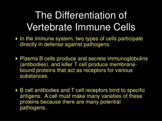
Download
1 / 11
140 likes | 323 Views
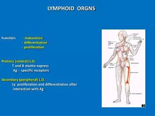
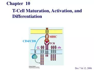
DIFFERENTIATION AND MATURATION OF T CELLS IN THE THYMUS. a. a. REGULATED T-CE L L DIFFERENTIATION. APC. CD4+ CD8+ TCR. preT- . Epithelial cell. immature T cell. pre T cell. ANTIGEN RECOGNIZING RECEPTOR. pro T c ell. SIGNALING RECEPTOR. NO ANTIGEN RECOGNIZING RECEPTOR.

E N D
a a REGULATED T-CELL DIFFERENTIATION APC CD4+CD8+ TCR preT- Epithelial cell immatureTcell pre Tcell ANTIGEN RECOGNIZING RECEPTOR pro T cell SIGNALING RECEPTOR NO ANTIGEN RECOGNIZING RECEPTOR
T- CELL DEVELOPMENT Lymphoid precursor NK cell c-kit/CD44 B B B B RAG-1/RAG-2 Pro-T Pro-B -rearrangement -rearrangement Pre-T L rearrangement Surrogate L H rearrangement Pre-T Pre-B T Selection clonal deletion Selection clonal deletion T T T Mature-T Mature-B
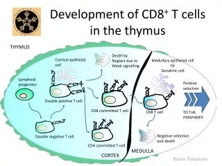
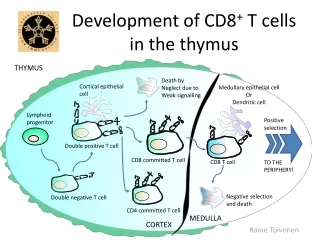
EVENTS OF T CELL DIFFERENTIATION IN THE THYMUS Pro-T IL-7-dependent proliferation Early pre-T Pre-Tα-chain Lck signal β rearrangement unsuccesful β-chain γδ T-cell No selection Late pre-T CD4+CD8+ α rearrangement CD4+CD8+ αβ NKT-cell unsuccesful α-chain no positive selection negative selection αβCD4+ αβCD8+ • Generation of NK cells • – no TCR • 2. Differentiation of γδ and αβ TCR carrying T cells • 3. Selection of αβ TCR • – positive selection • – negative selection • 4. Differentiation of CD4+ and CD8+ T cell lineages
SELECTION OF T LYMPHOCYTES IN THE THYMUS AICD – Activation Induced Apoptosis PERIPHERAL TOLERANCE UNDER THE CAPSULE • The primary T cell pool is biased to MHC-specificity (V genes) 1-2% for one allotype • Focusing the T cell pool to self MHC recognition (+) • Elimination of useless clones • Elimination of self agressive clones (-) • CENTRAL TOLERANCE • Focusing The T cell repertoire for recognition of non self • Individualized T cell repertoire is available in the periphery • CD4 and CD8 co-stimulatory molecules are involved in positive selection IL-7-dependent proliferation CORTEX CD4-CD8- DN β+preTα TCRαβ CD4+CD8+ DP TCR(-) sMHC+sP sMHC+fP fMHC+fP selection CORTEX/ MEDULLA NO – selection MEDULLA – AICD αβTCR αβTCR CD4+CD8+
SELECTION OF THE T CELL REPERTOIRE – CENTRAL TOLERANCE POSITIVE SELECTION – Thymic education (no instruction for specificity) Low avidity interaction of MHC - self peptide - TCR Thymic epithelial cells Self peptide composition and concentration (foreign peptides are not present) Low peptide dose induces positive selection – special ligands 80-90% of DN (CD4-CD8-) T cells is NOT positively selected PASSIVE CELL DEATH BY NEGLECTION NEGATIVE SELECTION – Central self tolerance High avidity of MHC - self peptide - TCR interaction Ubiquitous and abundant self antigens are present in the thymus High peptide dose induces negative selection Any thymic antigen presenting cell: epithelial cells, bone marrow-derived macrophages, dendritic cells THE GENERATION OF SELF MHC + FOREIGN PEPTIDE SPECIFIC T CELLS REQUIRES WEAK INTERACTION WITH SELF MHC + SELF PEPTIDE SELF RESTRICTED AND TOLERANT PERIPHERAL T CELL REPERTOIRE PHYSIOLOGICAL TRESHOLD
POSITIVE SELECTION OF DOUBLE POSITIVE (DP) T CELLS ALSO DIRECTS CD4 AND CD8 SINGLE POSITIVE (SP) T CELL COMMITMENT CD4+CD8+ CD4+CD8+ Thymic epithelial cell MHC-I + peptide complexes recruit CD8 MHC-II + peptide complexes recruit CD4 POSITIVE SELECTION FOR 3 – 4 DAYS, SUCCESSIVE α-GENE REARRANGEMENTS BARE LYMPHOCYTE SYNDROME (BLS) Lack of MHC class I – no CD8+ cells Lack of MHC class II – no CD4+ cells
PERIPHERAL TOLERANCE IMMUNE RESPONSES ARE NOT INITIATED IN THE PERIPHERY Normal tissue cells do not express MHC class II NO SIGNAL 1. for CD4+ Th activation Normal tissue cells do not express co-stimulatory molecules and do not produce T cell differentiating cytokines NO SIGNAL 2. for CD4+ Th activation Migration of naive T lymphocytes to normal tissues is limited Antigen presenting cells are not activated in normal tissues NO SIGNAL 3. for CD4+ Th activation PERIPHERAL TISSUES TOLERIZE THEMSELVES
MECHANISMS OF PERIPHERAL TOLERANCE ANERGY – Functional unresponsiveness, no IL-2 secretion SIGNAL 1Recognition of auto-antigen on tissue cell SIGNAL 2 No B7 and CD40 expression, no co-stimulation Tissue resident professional APC are not activatedSIGNAL 3Innate immunity is not activated No inflammation CLONAL DELETION – Activation induced cell death Requires persistant high antigen dose Fas – FasL interaction SUPPRESSION – Activity of other cells Cytokine-mediated balance Effector functions are inhibited by regulatory T cells CLONAL IGNORANCE No contact with the immune system Immunologically privileged sites Central nervous system, eye No recognition in the periphery
HOMEOSTASIS OF POSITIVE AND NEGATIVE SELECTION IN THE DEVELOPMENT OF THE AVAILABLE T LYMPHOCYTE REPERTOIRE Ratio of positive selection Homozygote Heterozygote Ratio of negative selection increases with the number of MHC genes Number of MHC molecules
a a APC APC APC APC APC APC CD8TCR CD8TCR CD8TCR CD4 TCR CD4 TCR CD4 TCR Ag Ag Ag T-CELL DIFFERENTIATION IN THE PERIPHERY Memory T-cell Activated T-cell Mature naiveT-cell