Download

1 / 67
670 likes | 1.04k Views
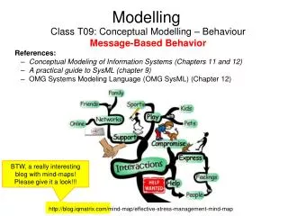
Modelling Cancer Growth. Philip K. Maini, Centre for Mathematical Biology, Mathematical Institute, Oxford. mutations. Approx 1mm in diameter. Nutrient required Hypoxic core TAF (tumour angiogenesis factors) Avascular tumour Vascular tumour Invasion

E N D
Modelling Cancer Growth PhilipK. Maini, Centre for Mathematical Biology, Mathematical Institute, Oxford
mutations Approx 1mm in diameter
Nutrient required Hypoxic core TAF (tumour angiogenesis factors) Avascular tumour Vascular tumour Invasion Tumour produces proteases – digest ECM Competition Normal environment: Tumour Normals Add H+ Gatenby & Gawlinski Gap
Acellular gap at the tumor-host interface in head and neck cancer
Hepatocytes Metastatic tumor
Tomas Alarcón (UCL)Helen Byrne (Nottingham)EU RTN (5th Framework): “Using mathematical modelling and computer simulation to improve cancer therapy”Alarcón, Byrne, Maini, J. Theor. Biol, 225, 257-274 (2003) Prog. Biophys & Mol. Biol., 85, 451-472 (2004) J. Theor. Biol, 229, 395-411 (2004) SIAM Multiscale Mod & Sim.3, 440-475 (2005) Ribba, Marron, Agur, Alarcon, Maini Bull. Math. Biol., 67 79-99 (2005)
Cancer Growth Tissue Level Signalling: (Tumour Angiogenesis Factors) Oxygen etc Cells: Intracellular: Cell cycle, Molecular elements Partial Differential Equations Automaton Elements Ordinary differential equations
Tumour Growth Response to mechanical stimuli Shrinkage (Pries et al 1988) First, work out distribution of 02 (nutrient) To do so, must consider vasculature: metabolic response R = radius = flow rate H = haematocrit Tw= WSS P = pressure (transmural) *Haematocrit Pries et al, 1994 *At a bifurcation: (rat mesentry) (Fung 1993)
Algorithm for structural adaptation Flow rate Prescribed 2. Given initial network configuration, compute flow rates through and pressure drops across each vessel using Kirchoff’s law. 3. Compute distribution of haematocrit. 4. Update radius of each vessel. 5. Compute viscosities (using H and R from 3 and 4 respectively). 6. Repeat until steady state reached.
____________O2 distribution____________ (adiabatic approx) P=O2 conc ΚN for normal cell Κc for cancer cell 0 o.w Nw= normal to vessel wall Ρb = О2level in blood P = permeability (at edge of domain, no flux
Automaton Rules 1. О2 distribution determined by BVP. 2. Cells attempt to divide at each time step. 3. Normal cell: if О2< threshold, cell dies О2 > threshold, cell attempts to divide Threshold = Ν1if more normal than cancer neighbours = ΝT2if more cancer than normal neighbours ΝT2> ΝT1 4. Cancer cell: if О2> threshold, cell attempts to divide Threshold = СT1if more cancer than normal neighbours = СT2if more normal than cancer neighbours С T2> СT1
5. Cancer cell: if O2< threshold cell becomes quiescent If it remains quiescent for a certain length of time, it dies.6. Cells are sinks of O27. If O2 level is such that a cell may divide, sample neighbourhood for space. If more than one available space, go to the one with largest O2 (Patel et al 2001). If no space, die (Kansal et al, 2000)
Cell Dynamics NxN automaton elements. State vector has 3 components: • Occupation: normal cell/cancer cell/vessel/empty • Cell status: proliferative/quiescent • Local О2 conc We assume, for simplicity, vessel structures does not evolve.
Conclusion • Environmental heterogeneity decreases cancer cell growth but may contribute to metastasis
Possible application Doxorubicin treatment of non-Hodgkin’s lymphoma (Ben Ribba, Zvia Agur, Tomas Alarcon, Philip Maini, K Marron) Structural adaptation – vessels surrounded by NHL leaky & unstable Nutrient diffusion -Drug pharmacokinetics in plasma pharmacodynamics [kills proliferating cells] tissue dynamics (adiabatic approx) AIM – Explore different protocols of treatment (presently a 21-day cycle is employed)
Cell-Cycle Dynamics Why? nutrient demand hypoxia-induced quiescence drugs work only on cells in a certain part of their cell cycle. Cell Cycle: Cyclin-dependent kinases (CDK) } cyclins } } In G1 CDK activity is low because its cyclin partners are missing At finish Cdhl (and Cdc 20) concs are high degrade cyclins. 2 families of proteins
Tyson & Novak • Model for G1/S transition
E2F – transcription factor Take Tyson and Novak model:incorporate inhibition by a – Kz term P27 conc in Cdhl oxygen Normals Growth regulation hypoxia [as m z ] Cancer Cells Hypothesis – growth regulation is lost
Simulations show decrease in Cdk • This is observed experimentally
Growth regulation of p27? Normals Cancer x Growth factors p27 If growth is arrested, p27 is upregulated
Response to hypoxia (low O2)Expts on mouse embryo fibroblasts: hypoxiaNormal cells G1arrest Does not occur with p27 null mutants
CONCLUSION x heterogeneities have a profound effect on tumour dynamics x effects of p27 – possible mechanism x efficiency of drug treatments Future Directions VEGF HIF-1 Elasticity