Download
1 / 53
570 likes | 925 Views
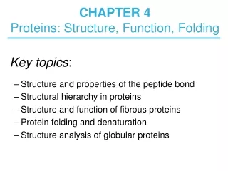
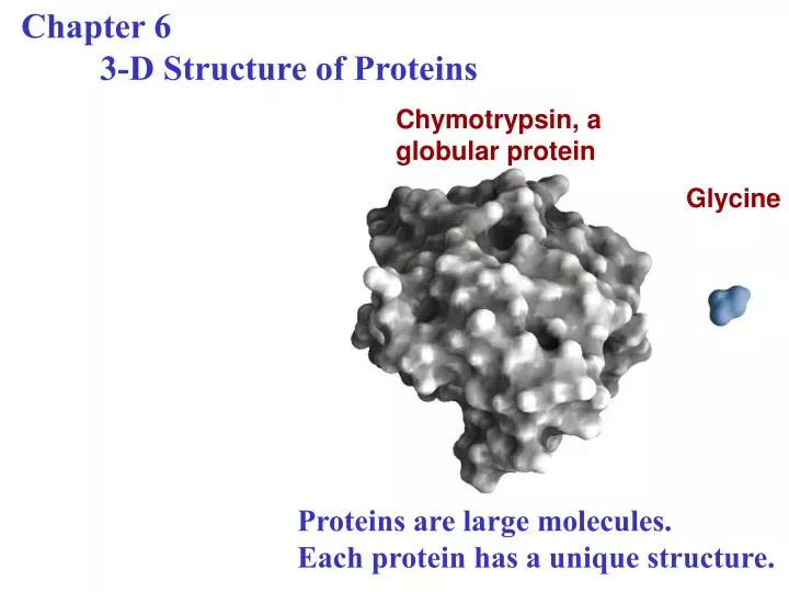
Chapter 6 3-D Structure of Proteins. Chymotrypsin, a globular protein. Glycine. Proteins are large molecules. Each protein has a unique structure. Overview of protein structure.

E N D
Chapter 6 3-D Structure of Proteins Chymotrypsin, a globular protein Glycine Proteins are large molecules. Each protein has a unique structure.
Overview of protein structure • For a protein numerous conformations are possible (without breaking a bond, by just rotation about single bonds) • However, a conformation existing under a given condition is the most stable thermodynamically having the lowest Gibbs free energy • Proteins in any of their functional folded conformations are called native protein
Peptide bond 1 3 2 Electric dipole
The peptide bond is rigid and planar C O C = N 1.27 Ao C N 1.47 Ao • 6 atoms lie in a single plane • Oxygen atom of the carbonyl group & the H of amide nitrogen are in trans • Bond between C and N has double bond character and is unable to rotate • Rotation is permitted between N – Ca and Ca– C • Polypeptide backbone: series of rigid planes with consecutive planes sharing a common point of rotation at Ca
Bond angles resulting from rotations at Ca C O Ca – C bond (psi) N – Ca bond (phi) • In the fully extended conformation f = y = 180o • y can have nay values between -180o to 180o • But many values are prohibited by steric interference
2 peptide bonds are in the same plane • f = y = 0o • This conformation is not allowed in proteins due to steric overlap between a-carbonyl oxygen and a-amino hydrogen atom
C Ramachandran Plot for L-Ala No steric overlap & Allowed conformation Permissible If little flexibility is allowed in the bond angle
Levels of structure in proteins Linking of aa Stable arrangement of aa give rise to structural patterns 3-dimensional folding of polypeptides Arrangement in space of 2 or more polypeptide subunits
Protein Secondary Structure • Refers to the local conformation of some part of polypeptide • Prominent secondary structures that are stable and occur in proteins are 1) a helix 2) b conformation
a helix • f = -60o • y = -45o to-50o • Each helical turn includes 3.6 aa • a helix found in all proteins is right handed Shows the hydrogen bonds Imaginary axis
a helix Looking down the longitudinal axis
Atoms in the center of the a helix are in very close contact
a helix • Form more readily • Helical structure is very stable • Because of internal hydrogen bonds • Between H attached to N of peptide linkage & the electronegative carbonyl O of the 4th aa on the N-terminal side of the peptide bond • Within the helix every peptide bond participates in such H bonding (except the last 4 amino acids that are close to the end of the helix)
AA sequence affects helix stability • For example, polypeptide with long block of Glu residues will not form an a helix at pH 7 • Bulk shape of Cys, Ser, Thr can destabilize the helix if they are close together in a chain • Proline introduces destabilizing kink in a helix • Gly occurs infrequently as it is very flexible & takes coiled structure different from a helix Interactions between R groups of aas 3 residues apart in a helix Arg (103) side chain Asp(100) side chain
Electric dipole of a peptidebond is transmitted through the hydrogen bonds along an a helix resulting in an overall helix dipole Negatively charged amino acids near the end of the amino terminal stabilize the positive charge of the helix electric dipole Positively charged amino acids near the end of the N-terminal destabilize the helix
The b conformation of the polypeptide chains Hydrogen bonds cross-links between adjacent chains The backbone of the polypeptide chain is extended into a zigzag structure Zigzag structures arranges side by side to form a structure resembling a series of pleats (called b sheet)
Most common structures of b turns (connecting elements) • Connects ends of 2 adjacent segments of an anti-parallel b sheet • 180o turn involving 4 amino acids • Gly and Pro occur in b turns (Gly at 3rd position) N H O C More common
Ramachandran plots for variety of structures Although theoretically possible, not observed in proteins
Relative probability that a given amino acid will occur in the 3 common types of secondary structure
Proteins are classified into 2 major groups: Globular proteins • Polypeptide folded into globular shape • Contain more than 1 type of secondary structure • Soluble in water Example: most enzymes Fibrous proteins • Polypeptide chain arranged in sheets or strands • Contain single type of secondary structure • Insoluble in water
Relationship between protein structure and biological function
Structure of hair (right-handed a helix) Higher order structures
Collagen a chain Structure of collagen Unique secondary structure distinct from a helix Repeating tripeptide sequence Gly-X-Pro or Gly-X-HyPro Adapts a left-handed helical structure with 3 residues per turn 3 separate a chains are supertwisted
Collagen with a distinct tertiary and quaternary structure • Collagen is • 35% Gly • 11% Ala • 21% pro and hyPro • Gelatin is derived from collagen Gly 3 stranded collagen superhelix
Structure of silk • Silk or spider web made up of the protein fibroin Layers of Antiparallel b sheet Small R groups allow close packing
Globular protein structures are compact and varied Human serum albumin 585 residues Actual size of the protein
Tertiary structure of sperm whale myoglobin 8 a helix Some bends which are b turns
Tertiary structure of sperm whale myoglobin Hydophobic residues (buried)
3-D structure of some small proteins Disulfide bonds a helix b sheet
Polypeptides (with more than few 100 aas) fold into 2 or more stable globular units called domains
Supersecondary structures (or motifs or folds) Stable arrangements of several elements of secondary structure and the connections between them Stable folding patterns in proteins
The quaternary structure of deoxyhemoglobin a subunits b subunits
Protein denaturation (loss of 3-D structure sufficient to cause loss of function)