Download
1 / 16
160 likes | 295 Views
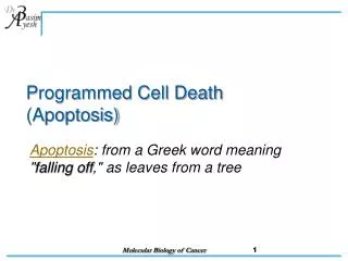
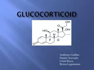
ROLE OF INTRACELLULAR CALCIUM IN GLUCOCORTICOID-EVOKED LYMPHOID CELL APOPTOSIS. Devin Morris California State University, Northridge. Stimuli/ Death signals. Release of cyctochrome c. Activation of caspase cascade. Apoptosis. Caspase 9. Apaf-1. Apoptosome formation. Effector

E N D
ROLE OF INTRACELLULAR CALCIUM IN GLUCOCORTICOID-EVOKED LYMPHOID CELL APOPTOSIS Devin Morris California State University, Northridge
Stimuli/ Death signals Release of cyctochrome c Activation of caspase cascade Apoptosis Caspase 9 Apaf-1 Apoptosome formation Effector Caspases Apoptosis
Cell Population Proliferation Cell Death Physiological roles of apoptosis • Development • Homeostasis • Disease
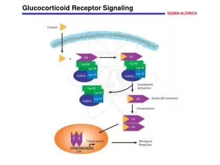
IPC 4 2 IPC R R R R R ABC R 3 1 coactivator complex R IPC interacting proteins basal trans. factors ligand receptor Glucocorticoid Pathway
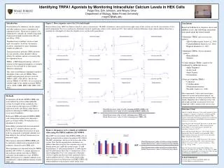
GC-induced Increase in Intracellular Calcium in GC-Susceptible CEM cells
EtOH 100 nM Dex 1mM Dex A B C 24 hours 4.4% 6% 14.1% D E F 48 hours 5% 24.8% 62.1% Time- & GC-Dose-Dependent Increase in [Ca+2]iin CEM-C7-14 Cells
Modulators of[Ca+2]i Levels Influence GC-Evoked Death of CEM Cells
EtOH 100 nM Dex 1mM Dex A B C Ethanol 4.4% 16.4% 35% D E F 20 mM EGTA 2% 12.4% 23.1% EGTA Suppresses GC-Evoked Increase in [Ca+2]i Levels in CEM-C7-14 Cells
Glucocorticoid Signaling Pathway Pathway Inhibitors Normal Pathway
Inhibition of Calmodulin Protects CEM-C7-14 Cells from GC-evoked Death
Inhibition of Calmodulin Kinase II Protects CEM-C7-14 Cells from GC-evoked Death
Inhibition of Calcineurin Protects CEM-C7-14 Cells from GC-evoked Death
Conclusions • GCs increase [Ca+2]i levels only in the GC-susceptible CEM-C7-14 cell line in a dose dependent manner; not in the GC-resistant sister cell line, CEM-C1-15. • Calcium chelation by either BAPTA or EGTA protected CEM-C7-14 cells from GC-evoked apoptosis, in conjunction with a reduction in the amount of free [Ca+2]i. • The calcium ionophore A23187 causes sensitization of CEM-C1-15 cells to GC-evoked apoptosis. • Inhibition of calmodulin, calmodulin kinase II or calcineurin, all intermediates in the calcium signaling pathway, impart varying degrees of protection to CEM-C7-14 cells from GC-evoked apoptosis. • Our data demonstrate a clear correlation between calcium signaling and GC-evoked apoptosis
Future Goals • Further studies will aim to understand Ca+2-dependent changes in gene regulation that contribute to apoptosis. Candidate genes such as the transcriptional repressor E4BP4, and its downstream targets are being studied. • Our ultimate goal is to understand the molecular pathway for apoptosis in T-lymphoid cells as well as in other physiologically relevant models for apoptosis, such as osteoblasts, keratinocytes and macrophages.
Acknowledgements Funded by grants from the NIH MBRS-SCORE Program, the CSUN Office of Graduate Studies, Research and International Programs, and the CSUN College of Science & Mathematics. • Dr. Rheem Medh • Saul Priceman • Dr. Carol Shubin • NASA CSUN/JPL Pair Program