Download
1 / 53
540 likes | 554 Views
Understand the significance of raised ICP, clinical features, and consequences of herniation. Dive into hydrocephalus causes, symptoms, and treatment options, including shunts. Learn about different hydrocephalic syndromes and the complex management of these conditions.

E N D
Disturbances of Intra-Cranial Pressure (ICP) and Hydrocephalus Andrew Danks Chairman of Neurosurgery, MMC
Significance of raised ICP depends clinical context • Fast vs Slow tempo • slow rise allows compensation • brain shift / moulding/atrophy • CSF shifts • even bone moulding / atrophy • Young child • splitting of sutures, head growth • can often allow compensation
Normal ICP • Normally 10-15 cm water in supine position • lower in young children • -5 to 0 cm water in standing position • at foramen of Munro (mid temple) • CSF actively secreted at 20 ml/hr or so • CSF resorbed at arachnoid villi in pressure-dependant mechanism • CSF pressure is the driver for head growth
Benign Intracranial Hypertension • Chronic raised ICP • up to 40 - 60 cm water • no hydrocephalus / brain distortion • normal brain function • may get headaches, • papilloedema, and visual loss • due to raised venous or CSF pressure
Clinical Features of Chronically Raised ICP • Symptoms : • headache • vomiting • impaired mentation, conscious state • Signs : • papilloedema : vision at risk • poor upgaze, 6th nerve palsy • impaired mentation, conscious state
Common causes of acute ICP • Severe head injury • Intracranial haematoma • Tumour / abscess • Infection - meningitis, encephalitis • Metabolic • Post operative swelling • Ischaemic strokes • Hydrocephalus • Sub-arachnoid haemorrhage
ICP Reflects : • Volume of contents / volume of cranium • contents = • brain • blood • CSF • pathology : tumour, haematoma, etc • oedema : intracellular / extracellular • normal ICP = 5 - 15 cm water, postural
CT and MRI show shapes of tissues, not pressure • Pressure can often be inferred • However, significant traps exist • in “acutely blocked shunt”, ventricles often are not dilated • some pts have slit ventricles when controlled • some pts do not dilate ventricles due to stiff walls, but pressure increased
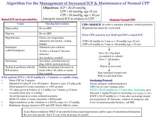
More important than ICP : • Cerebral perfusion pressure • CPP = Arterial pressure - ICP • accepted goal in ICU setting = 60 mmHg • Herniation • brain tissue forced between compartments • damage to this brain • further increased ICP
Consequences of herniation • Local damage in herniated tissue • infarction • vessel compression / traction • further oedema due to above • nerve damage due to pressure • 3rd nerve • CSF entrapment - more pressure
Acute trans-tentorial herniationunilateral • Medial temporal lobe forced into tentorial hiatus • Third nerve palsy, pupil first • mid-brain compression • ipsilateral • contralateral vs. opposite tentorial edge
Acute trans-tentorial herniationsymmetrical • Diencephalon symmetrically forced into tentorial hiatus • does NOT catch third nerve • symmetrical decline of conscious state with posturing etc. • pupils small not dilated
Treatment of Acutely Raised ICP • Diagnose and treat concurrently • ETT, hyperventilate and paralyse • Mannitol (1gm/kg) • CT • call neurosurgeon : specific treatments : • drain CSF in hydrocephalus • evacuate haematoma • dexamethasone for tumour oedema
Complicating factors in emergency neurosurgery • A • B • C • D dilutional : low sodium • E epilepsy • F fever : increases ICP, metabolism
Hydrocephalus • Acute vs. chronic • adult vs.infant • head size • non-communicating vs. communicating • former may be prone to rapid decline • LP dangerous in former, helpful in latter
Clinical Hydrocephalic Syndromes • acute hydrocephalus • headache, vomiting, drowsiness, declining consciousness, papilloedema • chronic hydrocephalus in child • big head, headache, papilloedema, false localizing signs • “normal Pressure” hydrocephalus • triad of gait apraxia, incontinence, dementia • may be sequel to SAH, meningitis,etc • compensated hydrocephalus • chronic ventriculomegaly, stabilized, asymptomatic • differential includes cerebral atrophy
Causes of hydrocephalus • Non-communicating : • tumour esp. in posterior fossa • aqueduct stenosis/blockage • Arnold - Chiari malformation • Communicating : • congenital • sludge in SA space : SAH, meningitis • blocked arachnoid villi • congenital
Obs. ZI…. Thomas ( 13 - 01 1992 ) • At 6 yrs : headaches, drowsiness, • rapid visual deterioration • 24-08-1998 : OD = 4/10 OG = 2/10 • CT and MRI : Craniopharyngioma • Hydrocephalus • 30-08-1998 : OD = 1/20 OG = 1/10 • VP Shunt • 02-09-1998 : OD = 3/10 OG = 2/10 • 03-09-1998 : Total resection of C. • 18-02-1999 : OD = 1/30 OG = 0
Modern Management of Hydrocephalus SHUNTS ‘ COST
Treatment of hydrocephalus • Treat cause if possible • eg remove tumour, treat meningitis • External ventricular drain if acute / infected • Lumbar puncture, IF COMMUNICATING • Ventriculo-peritoneal shunt - with valve • Other shunts : • V-Atrial, V-pleural, Lumbo- p • Endoscopic 3rd ventriculostomy • treatment of choice in aqueduct stenosis, 4th ventricle obstruction
Common problems with V-P shunts • Blockage - early or late • Infection - acute or delayed up to 6 months • Over-drainage • subdural hygroma/haematoma • slit ventricles, small head to due to chronic effects on head growth • headaches
Long term shunt survival Sigma Standard Delta 1.0 .8 Cumulative shunt survival .6 .4 p=.04 .2 0 1 2 3 4 5 6 Time (years) C. Sainte Rose
Has this person got a blocked shunt ? • Headache, drowsiness, N&V • GCS, eye movements, fundi • Does the valve pump and refill ? • Scan and compare • Very closely, slice by slice • The trap is interval decrease in vents after shunting, which may take 1 year, then later increase due to blockage • Catheter position, disconnection (XR series) • N/S Registrar
Intracerebral haemorrhage • Presentation – • Acute stroke, declining consciousness, seizure • Cause – • Aneurysm, AVM, trauma, hypertension • Surgical evacuation in minority • Young patients, larger lobar haematomas • Cerebellar haematomas • Not elderly, basal ganglia
Presentation of SAH • 5 ways
Presentation of SAH • sudden death • sudden LOC, recovering or persisting. • SUDDEN severe headache • meningeal signs / symptoms • lumbago, several days later
Diagnosis of SAH • 2 steps
Diagnosis of SAH • CT : acute blood is white • LP - if and only if CT is normal • best after 12 hours to allow xanthochromia • experienced operator - traumatic tap problematic