Download
1 / 43
430 likes | 512 Views
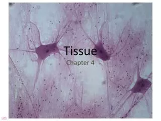
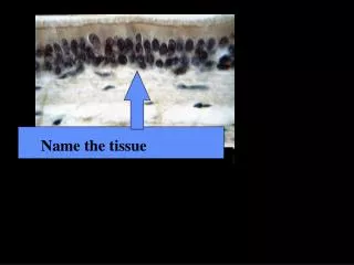
The tissue. Lab 3. Tissues. Groups of cells similar in structure and function The four types of tissues Epithelial Connective Muscle Nerve. Epithelial Tissue. Cellularity – composed almost entirely of cells

E N D
The tissue Lab 3
Tissues • Groups of cells similar in structure and function • The four types of tissues • Epithelial • Connective • Muscle • Nerve
Epithelial Tissue • Cellularity – composed almost entirely of cells • Special contacts – form continuous sheets held together by tight junctions and desmosomes • Polarity – apical and basal surfaces • Supported by connective tissue – reticular and basal laminae • Avascular but innervated – contains no blood vessels but supplied by nerve fibers • Regenerative – rapidly replaces lost cells by cell division
Classification of Epithelia • Simple or stratified Figure 4.1a
Classification of Epithelia • Squamous, cuboidal, or columnar Figure 4.1b
Epithelia: Simple Squamous Figure 4.2a
Epithelia: Simple Cuboidal • Single layer of cubelike cells with large, spherical central nuclei • Function in secretion and absorption • Present in kidney tubules, ducts and secretory portions of small glands, and ovary surface Figure 4.2b
Epithelia: Simple Columnar Figure 4.2c
Epithelia: Pseudostratified Columnar • Single layer of cells with different heights; some do not reach the free surface • Nuclei are seen at different layers • Function in secretion and propulsion of mucus • Present in the male sperm-carrying ducts (nonciliated) and trachea (ciliated) Figure 4.2d
Epithelia: Stratified Squamous • Thick membrane composed of several layers of cells • Function in protection of underlying areas subjected to abrasion • Forms the external part of the skin’s epidermis (keratinized cells), and linings of the esophagus, mouth, and vagina (nonkeratinized cells) Figure 4.2e
Epithelia: Transitional • Several cell layers, basal cells are cuboidal, surface cells are dome shaped • Stretches to permit the distension of the urinary bladder • Lines the urinary bladder, ureters, and part of the urethra Figure 4.2f
Epithelia: Glandular • A gland is one or more cells that makes and secretes an aqueous fluid • Classified by: • Site of product release – endocrine or exocrine • Relative number of cells forming the gland – unicellular or multicellular
Endocrine Glands • Ductless glands that produce hormones • Secretions include amino acids, proteins, glycoproteins, and steroids
Exocrine Glands • More numerous than endocrine glands • Secrete their products onto body surfaces (skin) or into body cavities • Examples include mucous, sweat, oil, and salivary glands • The only important unicellular gland is the goblet cell • Multicellular exocrine glands are composed of a duct and secretory unit
Structural Classification of Multicellular Exocrine Glands Figure 4.3a-d
Structural Classification of Multicellular Exocrine Glands Figure 4.3e-g
Connective Tissue • Found throughout the body; most abundant and widely distributed in primary tissues • Connective tissue proper • Cartilage • Bone • Blood
Connective Tissue Figure 4.5
Connective Tissue: Embryonic Figure 4.8a
Connective Tissue Proper: Loose Figure 4.8b
Connective Tissue Proper: Loose Figure 4.8c
Connective Tissue Proper: Loose Figure 4.8d
Connective Tissue Proper: Dense Regular Figure 4.8e
Connective Tissue Proper: Dense Regular Figure 4.8f
Connective Tissue: Hyaline Cartilage Figure 4.8g
Connective Tissue: Elastic Cartilage • Similar to hyaline cartilage but with more elastic fibers • Maintains shape and structure while allowing flexibility • Supports external ear (pinna) and the epiglottis Figure 4.8h
Connective Tissue: Fibrocartilage Cartilage • Matrix similar to hyaline cartilage but less firm with thick collagen fibers • Provides tensile strength and absorbs compression shock • Found in intervertebral discs, the pubic symphysis, and in discs of the knee joint Figure 4.8i
Connective Tissue: Bone (Osseous Tissue) Figure 4.8j
COMPACT BONE it forms the walls & outer surfaces of the bones • Osteon- functional unit of the compact bone It is thickest where angular stress is applied . Lamella- the layers of the matrix that makes the bone ( thin plate) • Central canals (Harvesian canals) F-they contain blood vessels that carry blood to & from the Osteon Perforating canals (Canals of Volkmann) They extend perpendicular to the surface F- they connect central canals of adjacent osteons to each other • Lacuna- thin holes that contains an osteocyte It is a pocket sandwiched between layers of matrix Canaliculus- narrow pathway that penetrates the lamellae Function- to deliver nutrients and removal of waste products to and from the osteocytes
COMPACT BONE (MARTINI PG. 186)
SPONGY BONE • IT MAKES THE INNER LAYER(S) OF BONES • IT IS FORMED BY TRABECULAE (BONY BARS OR PLATES) • IT HAS SPACES FOR BLOOD CELL FORMATION • TRABECULAE CONTAINS LACUNA WITH OSTEOCYTES DR. ALFONSO A PINO
Connective Tissue: Blood Figure 4.8k
Epithelial Membranes • Cutaneous – skin Figure 4.9a
Epithelial Membranes • Mucous – lines body cavities open to the exterior (e.g., digestive and respiratory tracts) • Serous – moist membranes found in closed ventral body cavity Figure 4.9b
Epithelial Membranes Figure 4.9c
Nervous Tissue Figure 4.10
Muscle Tissue: Skeletal • Long, cylindrical, multinucleate cells with obvious striations • Initiates and controls voluntary movement • Found in skeletal muscles that attach to bones or skin Figure 4.11a
Muscle Tissue: Cardiac • Branching, striated, uninucleate cells interdigitating at intercalated discs • Propels blood into the circulation • Found in the walls of the heart Figure 4.11b
Muscle Tissue: Smooth Figure 4.11c
Developmental Aspects • Primary germ layers: ectoderm, mesoderm, and endoderm • Three layers of cells formed early in embryonic development • Specialize to form the four primary tissues • Nerve tissue arises from ectoderm
Developmental Aspects • Muscle, connective tissue, endothelium, and mesothelium arise from mesoderm • Most mucosae arise from endoderm • Epithelial tissues arise from all three germ layers
Developmental Aspects Figure 4.13