Download
1 / 44
470 likes | 734 Views
Tissue. The Living Fabric. Section 1 . Pages 118-124. Epithelial Tissue. Epithelial tissue (epithelium) Sheet of cells that covers a body surface or lines a body cavity. Two types: Covering and lining epithelium Outer layer of skin, lines open cavities Glandular epithelium

E N D
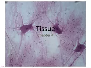
Tissue The Living Fabric
Section 1 Pages 118-124
Epithelial Tissue • Epithelial tissue (epithelium) • Sheet of cells that covers a body surface or lines a body cavity. • Two types: • Covering and lining epithelium • Outer layer of skin, lines open cavities • Glandular epithelium • Fashions the glands of the body
Epithelial Tissue • Epithelium has many functions: • Protections • Absorption • Filtration • Excretion • Secretion • Sensory reception
Classification of Epithelia • Each epithelium is given two names • First - # of layers • Simple and stratified • Simple epithelia – single cell layer (found where absorbtion and filtration occur • Stratified epithelia – two or more layers (high abrasion areas)
Classification of Epithelia • Second name – shape of cells • Three common shapes • Squamous cells – flattened and scalelike • Cuboidal cells – boxlike • Columnar cells - tall and column shaped
Simple Epithelia • Simple Squamous • Flattened laterally, cytoplasm is sparse • Look like a fried egg • Allows passage of materials by diffusion and filtration • Areas where protection is not important • Kidneys, air sacs of lungs, lining of heart • Two names that reflect their location • Endothelium – ‘inner covering’ • Mesothelium – covering organs
Simple Cuboidal Epithelium • Consists of a single layer of cells as tall as they are wide, spherical central nuclei • Functions: • Secretion • Absorption • Location • Ovary surface, ducts and secretory protions of small glands
Simple Columnar Epithelium • Single layer of tall closely packed cells, round/oval nuclei • Some contain cilia which help move substances through pathway • Function: • Absorption, secretion of mucus • Location: • Lines most of the digestive tract, gallbladder
Stratified Epithelia • Two or more layers of cells • More durable • Regenerate from below • Role is protection
Stratified Epithelia • Stratified Squamous Epithelium • Thick membrane composed of several layers • Function: • Protects underlying tissues in areas subjected to abrasion • Locations: • Moist linings of mouth and esophagus
Stratified Epithelia • Stratified Cuboidal and Columnar Epithelia • Cuboildal • Quite rare in the body • Mostly found in ducts of larger glands • Columnar • Small amounts are found in pharynx, and lining of some glandular ducts
Connective Tissue • Found everywhere in the body • Most abundent and widely distributed of the primary tissues • Four main classes • Connective tissue proper • Catilage • Bone tissue • blood
Connective Tissue • Functions: • Binding and support • Protection • Insulation • Transportation of substances within the body
Common Characteristics • Despite their diverse functions, they have many common characteristics • Common origin • All connective tissue comes from mesenchyme (embryotic tissue) • Degrees of vascularity • Have many degrees of vascularity (contain vessels) • Extracellular Matrix • Seperates the living cells of the tissue • Because of matrix connective tissue can bear weight
Structural Elements of Connective Tissue • Connective Tissues have 3 main elements: • Ground substance • Fibers • Cells
Ground Substance • Unstructured material that fills the space between the cells and contains the fibers • Composed of: • Interstitial fluid • Cell adhesion proteins • Acts like glue, helps attach themselves to matrix elements • Proteoglycans • Holds large amounts of fluid and functions as a molecular sieve, or medium for nutrients to diffuse between blood capliaries and cells
Fibers • Provide support • Three main types of fibers: • Collagen • Elastic • Reticular fibers
Fibers • Collagen fibers • By far the strongest and most abundant • Constructed mainly of collagen • Secreted into the extracellular space • Cross-linked fibrils • Because of this cross-linked pattern they are very strong • Have a glistening white appearance – also called white fibers
Elastic Fibers • Long, thin fibers that form branching networks in the extracellular matrix • Contain rubber-like protein elastin • Allows them to stretch and recoil • Found where elasticity is needed • Skin, lungs, and blood vessel walls • Sometimes called yellow fibers
Reticular Fibers • Short, fine, collagenous fibers • Branch extensively, forming delicate networks that surround small blood vessels and support soft tissue
Cells • Each connective tissue has a fundamental cell type • Blast – ‘bud’ or ‘sprout’, means ‘forming’ • Primary blast cell types by connective tissue class are: • Connective tissue proper (fibroblast) • Cartilage (Chondroblast) • Bone (osteoblast) • Blood (hematopoietic stem cell) • Is no located in its tissue (blood) • These cells make the matrix of their connective tissue.
Connective Tissue Proper • Has two subclasses: • Loose connective tissues • Dense connective tissues • Except for bone and blood, all mature connective tissues belong to this class
Loose Connective Tissue • Areolar Connective Tissue • Gel-like matrix with three fiber types • Fibroblasts, macrophages, mast cells, and some white blood cells • Function • Wraps and cushions organs • Plays important role in inflammation (holds fluids) • Location • Widely distributed under epithelia of body • Surrounds capillaries
Loose Connective Tissue • Adipose (fat) tissue • Closely packed fat cells • Have nucleus pushed to side by large fat droplet • Function: • Provides reserve food fuel • Insulates against heat loss • Supports and protects glands • Location: • Under skin • Around kidneys and eyeballs • Within abdomen and breasts
Loose Connective Tissue • Reticular Connective Tissue • Network of reticular fibers in a typical loose ground substance; lie on the network • Function: • Fibers from soft internal skeleton that supports other cell types of cells including white blood cells • Location: • Lymphoid organs
Pictures Areolar Tissue Adipose Tissue Reticular Tissue
Cartilage • Stands up to both tension and compression • Has qualities imtermediate between dense connective tissue and bone • Tough but flexible • Made up of 80% water • Gets nutrients from blood vessels in membranes • Three varieties of cartilage: • Hyaline cartilage • Elastic cartilage • fibrocartilage
Hyaline Cartilage • Most abundent • Contains large amounts of collagen, not apparent in matrix • Appears glassy (hyalin = glass) • Provides support with some pliability • Absorb compression at joints • Supports tip of nose, connects ribs to sternum • Supports respiratory pathways
Elastic Cartilage • Description: • Similar to hyaline cartilage, but more elastic fibers in matrix • Function: • Maintains the shape of structure • Allows great flexibility • Location: • Supports the external ear
Fibrocartilage • Description: • Less firm than hyaline cartilage • Thick collagen fibers • Function: • Tensile strength with the ability to absorb commpressive shock • Location: • Intervertebral discs • Discs of knees
Pictures Fibrocartilage Hyaline Cartilage Elastic Cartilage
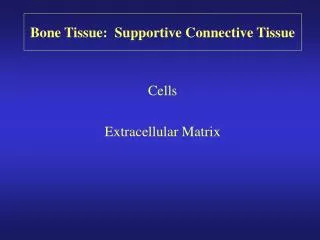
Bone • Rocklike hardness osseous tissue, has an exceptional ability to support and protect body structures • Provide cavity for fat storage • Synthesis of blood cells • Inorganic calcium salts • Seen as closely packed structural units called osteons, form rings
Blood • Blood • Fluid within blood vessels • Does not connect things to give mechanical support • Classified as connective tissue because it developes from mesynchyme and consists of blood cells surrounded by a nonliving matrix called blood plasma • Majority is red blood cells • ‘fibers’ are soluble protein molecules that precipitate, forming large fiberlike structures during clotting • Transport vehicle • Carries nutrients, waste and respiratory gases throughout body
Pictures Red Blood Tissue Bone Tissue White Blood Tissue
Nervous Tissue • Main component of nervous system • Brain, spinal cord, nerves • Neurons • Highly specialized nerve cells that generate and conduct nerve impulses • Branching cells – cell processes • Transmit electrical eignals from sensory receptors to effectors which control their activity
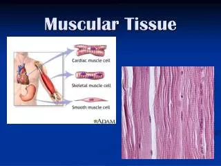
Muscle Tissue • Responsible for most types of body movements • Three kinds of muscle tissue: • Skeletal • Cardiac • Smooth
Skeletal Muscle • Attached to bones of the skeleton • Form the flesh of the body • When they contract they pull on bone or skin to produce movement • Long, cylindrical, many nuclei, obvious striations • Voluntary movement, facial expression, voluntary control
Cardiac Muscle • Found only in the wall of the heart • Helps propel blood throughout body • Branching, striated, generally uninucleate
Smooth Muscle • No visible striations • Walls of hallow organs • Digestive and urinary tract organs • Works to squeeze substances through these organs by alternated contracting and relaxing • Both smooth and cardiac muscles are involuntary muscle – we do not think about it when it is working
Steps of Tissue Repair • Repair occurs in two major ways: • Regeneration • Replacement of destroyed tissue • Fibrosis • Proliferation of fibrous connective tissue called scar tissue • Which of these occurs is based on: • Type of tissue damaged • Severity of the injury