Download
1 / 21
210 likes | 393 Views
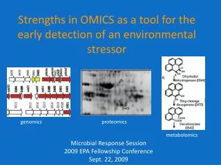
1 st Annual Unither Nanomedical and Telemedical Technology Conference. AFM force spectroscopy as a nanotool for early detection of misfolded protein. Alexey V. Krasnoslobodtsev, PhD. Outline. Misfolding (conformational) diseases – background.

E N D
1st Annual Unither Nanomedical and Telemedical Technology Conference AFM force spectroscopy as a nanotool for early detection of misfolded protein. Alexey V. Krasnoslobodtsev, PhD
Outline • Misfolding (conformational) diseases – background. • Single molecule approach (Force spectroscopy) to study misfolding phenomenon. • Force spectroscopy - advantages and applications. • Beyond measuring forces of intermolecular interactions – Dynamic Force Spectroscopy.
Protein aggregation Disease Protein fibrils Protein folding, misfolding and aggregation Environmental Stress Misfolded protein Chemical Stress Chaperones Native folded protein Generic Perturbations Pathophysiological Stress
Protein Misfolding (Conformational) Diseases Many human diseases are now recognized to be conformational diseases associated with misfolding of the proteins and their consequent aggregation. Alzheimer’s Parkinson’s Lewy bodies Plaques and tangles Prion amyloid plaques Huntington’s intranuclear inclusions Amyotrophic lateral sclerosis aggregates These diseases include neurodegenerative disorders such as Alzheimer’s, Parkinson’s disease, Huntington’s and prion diseases characterized by deposition of aggregates in Central Nervous System (CNS). • Misfolded proteins are prone to aggregation • Misfolded proteins and aggregates cause molecular stress and interfere with cellular function Claudio Soto, 2003
Mechanism of aggregation Normally folded protein Misfolded protein Oligomers Stress (environmental) induced misfolding generates “sticky” aggregation prone conformation Normally folded protein interacts with misfolded protein Cycle multiplies copies of misfolded (diseased) proteins Goal - looking at the first stage of aggregation (dimerization) at a single molecule level Large aggregates and fibrils
Possible therapeutic interventions for protein misfolding diseases Skovronovsky D.M., et al., 2006, Annu. Rev. Pathol. Dis., 1:151-70
Therapeutic approaches to misfolding diseases Expression of the protein Small molecules that bind to specific regions of the misfolded protein and stabilize it. Chemical (pharmacological) Chaperones Protein misfolding Prevent aggregation of misfolded proteins Aggregation Loss of neuronal function and cell death Neurodegeneration
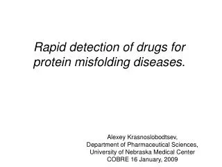
Rationale Despite the crucial importance of protein misfolding and abnormal interactions, very little is currently known about the molecular mechanism underlying these processes. Rationale: A clear understanding of the molecular mechanisms of misfolding and aggregation will facilitate rational approaches to prevent protein misfolding mediated pathologies. Initial stages of misfolding and aggregation are very dynamic. High-resolution methods such as x-ray crystallography, NMR, electron microscopy, and AFM imaging have provided some information regarding the secondary structure of aggregated proteins and morphologies of self-assembled aggregates. But they are unable to characterize transient intermediates that can not be detected by these bulk methods. We propose a novel method for identification and characterization of misfolded aggregation prone states of a protein as well as conditions favoring or disfavoring aggregation (misfolding). Single molecule force spectroscopy is capable of detecting interactions between transient species.
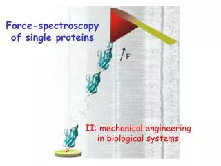
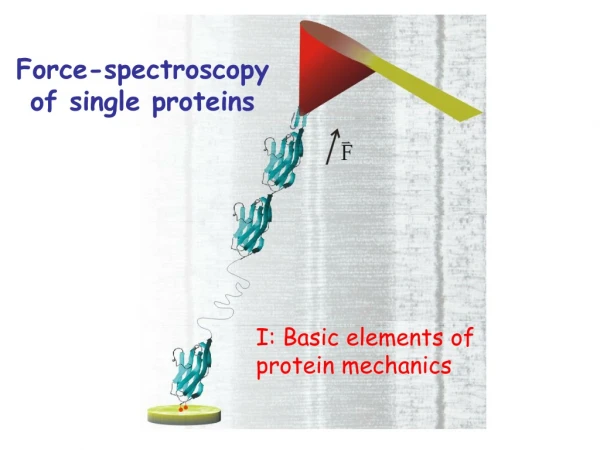
Dimerization of misfolded proteins is the very first step in aggregation process. Probing interactions between individual molecules by AFM force spectroscopy Force AFM force spectroscopy allows studying: Distance • Binding strengths - measures forces of interactions between individual molecules.
AFM force spectroscopy 2) Contact of the tip with sample surface Approaching 1) 4) 3) 5) 2 Rupture event Rupture force 3 4 5 Tip retraction Bond rupture Stretching the linkers
Model system- 7 aa peptide from Sup35 yeast prion 1 122 253 685 7GNNQQNY13 Misfolding – exposing “hot” regions Aggregation “Hot” regions are short stretches of peptide sequences. Alzheimer’s: amyloid-beta peptide 1-40(42) -> Aβ16-22is responsible for aggregation. Huntington’s: polyQ (>40) -> elementary Q7 shows maximal kinetics of aggregation. Parkinson’s: α-synuclein -> 12 aa regions is the core domain for aggregation. Prion diseases: short peptide from Sup35 yeast prion • A seven amino acid sequence within the N-terminal domain is responsible for the aggregation of the whole Sup35 protein • Sequence: GNNQQNY Nelson, R.R., Sawaya, M.R., Balbirnie, M., Madsen, A.Ø., Riekel, C., Grothe, R., Eisenberg, D. 2005. Structure of the cross-β spine of amyloid-like fibrils. Nature. Vol. 435, No. 9, 773-778.
Environmental Stress Misfolded 1 pH 2 Misfolded 2 pH 3.7 Misfolded 3 pH 5.6 Sup35 Aggregation at different pHs (Environmental Stress) Morphology of aggregation – different misfolding states that have different strength of interactions?
Parallel circular dichroism (CD) measurements performed for Aβ peptide revealed that the decrease in pH is accompanied by a sharp conformational transition from a random coil at neutral pH to the more ordered, predominantly β-sheet, structure at low pH. Importantly, the pH ranges for these conformational transitions coincide with those of pulling forces changes detected by AFM. In addition, protein self-assembly into filamentous aggregates studied by AFM imaging was shown to be facilitated at pH values corresponding to the maximum of pulling forces. Overall, these results indicate that proteins at acidic pH undergo structural transition into conformations responsible for the dramatic increase in interprotein interaction and promoting the formation of protein aggregates. AFM force spectroscopy – nanotool for detection of misfolded state. Amyloid -β peptide
AFM force spectroscopy -High throughput screening machine for detecting efficient therapeutic agents Drug #2 is the best candidate for the development of effective therapeutic agents Control Drug #1 Drug #2 Drug #3 Force of intermolecular interactions
Challenges Peptide • Robust system (for continuous measurements) We have recently developed surface chemistry which allows simple and reliable covalent attachment of biomolecules to the surfaces (AFM tip and mica). Peptide-SH • Automated exchange of buffers containing drugs of interest. • Automated data analysis.
Beyond Force SpectroscopyDynamic Force Spectroscopy (DFS) measurements DFS – measures kinetic parameters of dissociation reaction 2 1 r – pulling velocity (loading rate) F1 < F2
Dynamic Force Spectroscopy PP P + P koff Loading Rate ΔG‡ xβ Loading rate Distance to transition state Dissociation rate constant Force ln r
Dynamic Force Spectroscopy GNNQQNY – Sup35 yeast prion A dynamic force spectrum at pH=2.0 reveals two linear regimes distinguishable by differences in slopes. This is usually attributed to a molecular dissociation of a complex that involves overcoming of more than one activation barrier.
These data suggest that the ability of misfolded protein to form a stable dimer is a unique property of these conformational states for proteins suggesting a possible explanation for the phenomenon of the protein self-assembly into nanoaggregates. Dynamic Force Spectroscopy k1off k2off 0.2 Å 3.5 Å • Two barriers in the energy profile: • Inner (second fit) and outer (first fit) activation barriers • The estimated positions of inner and outer barriers are 0.2 and 3.5 Å. • The off rates are 286 and 0.9 s-1. • Estimated lifetime of a dimer is 1.1 s which is much longer than nano/microsecond conformational dynamics of a monomer.
Summary • Novel nanoprobing approach to study initial steps of misfolding and aggregation is proposed on the basis of AFM force spectroscopy operating on a single molecule level. • There is an intimate relationship between aggregation propensity (protein misfolding) and strength of interprotein interactions. • Force spectroscopy allows to study the mechanism of early dynamic events in the aggregation process which is not accessible by any other available method. • A dimer formed by two misfolded peptides is very stable as compared to monomer conformational dynamics providing the explanation for the phenomenon of the protein self-assembly into nanoaggregates.
Acknowledgements • Yuri L. Lyubchenko, Ph.D., D. Sc. • Lab Members: • Luda Shlyakhtenko, Ph.D. • Alex Portillo • Jamie Gilmore • Junping Yu, Ph.D. • Mikhail Karymov, Ph.D. • Shane Lippold • Nina Filenko, Ph.D. • Igor Nazarov, Ph.D. • Alexander Lushnikov, Ph.D Supported by NIH and Nebraska Research Initiative (NRI) grants to YLL