Download
1 / 51
620 likes | 1.05k Views
Thyroid Disorders. Thyroid hormones include: Thyroxine T4 and Triiodothyronine T3 THEIR SYNTHESIS AND RELEASE DEPEND ON TSH Feedback inhibition regulate pituitary production of TSH. GOITER. Is a general term for enlargement of the thyroid CAUSES: Physiologic enlargement

E N D
Thyroid hormones include: Thyroxine T4 and Triiodothyronine T3 THEIR SYNTHESIS AND RELEASE DEPEND ON TSH Feedback inhibition regulate pituitary production of TSH
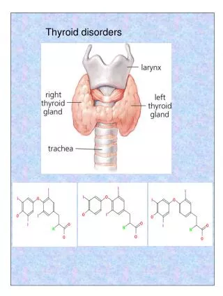
GOITER Is a general term for enlargement of the thyroid CAUSES: Physiologic enlargement Iodine deficiency Hashimoto thyroiditis Goitrogens Enzyme defficiency
SIMPLE GOITER: Goiter without thyroid hormone dysfunction TOXIC GOITER: Goiter associated with hyperthyroidism ENDEMIC GOITER: Occuring with high frequency in iodine-deficient areas NODULAR GOITER: Irregular enlargement of the thyroid, resulting in nodule formation
Hypothyroidism • Etiology: • Primary: • Hashimoto’s thyroiditis with or without goitre • Radioactive iodine therapy for Graves’ disease • Subtotal thyroidectomy for Graves’ disease or nodular goitre • Excessive iodine intake • Subacute thyroiditis • Rare causes • Iodide deficiency • Goitrogens such as lithium; antithyroid drug therapy • Inborn errors of thyroid hormone synthesis • Secondary: Hypopituitarism • Tertiary: Hypothalamic dysfunction (rare) • Peripheral resistance to the action of thyroid hormone
Manifested as myxedema in adults and cretinism in children MYXEDEMA: More common in women than men CAUSES: Therapy for hyperthyroidism Hashimoto thyroiditis Iodine deficiency Primary idiopathic myxedema
Clinical features • Weight gain • Cold intolerance • Rough, dry skin • Puffy face and hands • Hoarse, husky voice • Yellowish color of skin due to reduced conversion of carotene to vitamin A • Respiratory failure • Menorrhagia, infertility, hyper- prolactinemia • Anemia: • CVS: Bradycardia • Neuromuscular system: • Slow reflexes • CNS symptoms: • Fatigue, lethargy, depression • Inability to concentrate
CRETINISM: Causes: Iodine deficiency Enzyme deficiency Maldevelopment Failure to descend
CLINICALLY: Severe mental retardation Impairment of physical growth Protuberant abdomen
Hypothyroidism • Diagnosis: • A iFT4 and hTSH is diagnostic of primary hypothyroidism • Serum T3 levels are variable (maybe in normal range) • +ve test for thyroid autoantibodies PLUS an enlarged thyroid gland suggest Hashimoto’s thyroiditis • TRH test may be done to differentiate pituitary from hypothalamic disease. Absence of TSH response to TRH indicates pituitary deficiency • MRI of brain is indicated if pituitary or hypothalamic disease is suspected. Need to look for other pituitary deficiencies.
Hashimoto’s Thyroiditis • Hashimoto’s thyroiditis is a commom cause of hypothyroidism and goitre especially in children and young adults. • It is an autoimmune disease that involves heavy infiltration of lymphocytes that totally destroys normal thyroidal architecture • It is familial and may be associated with other autoimmune diseases such as pernicious anemia, adrenocortical insufficiency, idiopathic hypoparathyroidism, and vitiligo.
Hashimoto’s Thyroiditis • Symptoms & Signs: • Usually presents with goitre in a patient who is euthyroid or has mild hypothyroidism • Sex distribution: four females to one male • The process is painless • Older patients may present with severe hypothyroidism with only a small, firm atrophic thyroid gland • Transient symptoms of thyrotoxicosis can occur during periods of hashitoxicosis (spontaneously resolving hyperthyroidism) • Lab: • Normal or low thyroid hormone levels, and if low, TSH is elevated • High Tg Ab and/or TPO Ab titres • FNA bx reveals a large infiltration of lymphocytes
Management of Hypothyroidism • Start patient on L-thyroxine . • Hypothyroidism during pregnancy: • Check TFT every month. L-thyroxine dose requirement tends to go up as the pregnancy progresses.
Myxedema Coma • Medical emergency, end stage of untreated hypothyroidism • Characterized by progressive weakness, hypothermia, hypoventilation, hypoglycemia, hyponatremia, shock, and death • The patient (or a family member) may recall previous thyroid disease, radioiodine therapy, or thyroidectomy • iHR and marked hypothermia (as low as 24C) • The pt is usually an obese elderly woman with yellowish skin, a hoarse voice, thin hair, puffy eyes, ileus, and slow reflexes. An anterior neck scar may be present. Scanty pubic or axillary hair with pituitary myxedema • May be ppt by HF, pneumonia, excessive fluid administration, narcotics
Graves’ Disease • Most common form of thyrotoxicosis • May occur at any age but mostly from 20-40 • 5 times more common in females than in males • Syndrome consists of one or more of the following: • Thyrotoxicosis • Goitre • Opthalmopathy (exopthalmos) and • Dermopathy (pretibial myxedema) • It is an autoimmune disease of unknown cause • 15% of pts with Graves’ have a close relative with the same disorder
Graves’ Disease • Pathogenesis: • T lymphocytes become sensitized to Ag within the thyroid gland and stimulate B lymphocytes to synthesize Ab to these Ag • One such Ab is the TSH-R Ab(stim), which stimulates thyroid cell growth and function • The opthalmopathy and dermopathy associated with Graves’ may involve lymphocyte cytokine stimulation of fibroblasts in these locations causing an inflammatory response that leads to edema, lymphocytic infiltration, and glycosaminoglycans deposition • The tachycardia, tremor, sweating, lid lag, and stare in Graves’ is due to hyperreactivity to catecholamines and not due to increased levels of circulating catecholamines
Graves’ Disease • Clinical features: • I Eye features: Proptosis Extraocular muscle involvement Corneal involvement Sight loss (optic nerve involvement)
Graves’ Disease • Clinical features: • II Goitre: • Diffuse enlargement of thyroid • Bruit may be present • III Thyroid dermopathy (pretibial myxedema): • Thickening of the skin especially over the lower tibia • The dermopathy may involve the entire leg and may extend onto the feet • Skin cannot be picked up between the fingers • Rare, occurs in 2-3% of patients • Usually associated with opthalmopathy and very hTSH-R Ab
Graves’ Disease • Clinical features: • IV Heat intolerance • V Cardiovascular: • Palpitation, Atrial fibrillation • CHF, dyspnea, angina • VI Gastrointestinal: • Weight loss, happetite • Diarrhea • VII Reproductive: • amenorrhea, oligo- menorrhea, infertility • Gynecomastia • VIII Bone: • Osteoporosis • Thyroid acropachy • IX Neuromuscular: • Nervousness, tremor • Emotional lability • Proximal myopathy • X Skin: • Pruritus • Vitiligo, hair thinning • Palmar erythema • Spider nevi
Graves’ Disease • Diagnosis: • Low TSH, High FT4 and/or FT3 • If eye signs are present, the diagnosis of Graves’ disease can be made without further tests • If eye signs are absent and the patient is hyperthyroid with or without a goitre, a radioiodine uptake test should be done. • Radioiodine uptake and scan: • Scan shows diffuse uptake • Uptake is increased • TSH-R Ab (stim) is specific for Graves’ disease.
Treatment of Grave’s Disease • There are 3 treatment options: • Medical therapy • Surgical therapy • Radioactive iodine therapy
Toxic Adenoma (Plummer’s Disease) • This is a functioning thyroid adenoma • Typical pt is an older person (usually > 40) who has noted recent growth of a long-standing thyroid nodule • Thyrotoxic symptoms are present but no infiltrative opthalmopathy. PE reveals a nodule on one side • Lab: low TSH, high T3, slightly high T4 • Thyroid scan reveals “hot” nodule with suppressed uptake in contralateral lobe • Toxic adenomas are almost always follicular adenomas and almost never malignant • Treatment: same as for Grave’s disease
Toxic Multinodular Goitre • Usually occurs in older pts with long-standing MNG • PE reveals a MNG that may be small or quite large and may even extend substernally • RAI scan reveals multiple functioning nodules in the gland or patchy distribution of RAI • Hyperthyroidism in pts with MNG can often be ppt by iodide intake “jodbasedow phenomenon”. • Amiodarone can also ppt hyperthyroidism in pts with MNG • Treatment: Same as for Grave’s disease. Surgery is preferred.
Other Forms of Thyrotoxicosis • Thyrotoxicosis Factitia: • Due to ingestion of excessive amounts of thyroxine • RAI uptake is nil and serum thyroglobulin is low • Struma Ovarii: • Teratoma of the ovary with thyroid tissue that becomes hyperactive • No goitre or eye signs. RAI uptake in neck is nil but body scan reveals uptake of RAI in the pelvis. • Hydatidiform mole: • Chorionic gonadotropin is produced which has intrinsic TSH-like activity. • TSH-secreting pituitary adenoma: • FT4 & FT3 is elevated but TSH is normal or elevated • Visual field examination may reveal temporal defects, and CT or MRI of the sella usually reveals a pituitary tumour.
Thyroid storm (Thyrotoxic crisis) • Usually occurs in a severely hyperthyroid patient caused by a precipitating event such as: • Infection • Surgical stress • Stopping antithyroid medication in Graves’ disease • Clinical clues • fever hyperthermia • marked anxiety or agitation coma • Anorexia • tachycardia tachyarrhythmias • pulmonary edema/cardiac failure • hypotension shock • confusion
Thyroid storm (Thyrotoxic crisis) • Initiate prompt therapy after free T4, free T3, and TSH drawn without waiting for laboratory confirmation. • Therapy • 1. General measures: • Fluids, electrolytes and vasopressor agents should be used as indicated • A cooling blanket and acetaminophen can be used to treat the pyrexia • Propranolol for beta–adrenergic blockade and in addition causesdecreased peripheral conversion of T4T3 but watch for CHF. • The IV dose is 1 mg/min until adequate beta-blockade has been achieved. Concurrently, propranolol is given orally or via NG tube at a dose of 60 to 80 mg q4h
Thyroid storm (Thyrotoxic crisis) • Therapy • 2. Specific Measures: • anti-thyroid drug of choice • Dexamethasone • 3. Identify and treat precipitating factor.
Nontoxic Goitre • Enlargement of the thyroid gland from TSH stimulation which in turn results from inadequate thyroid hormone synthesis • Etiology: • Iodine deficiency • Goitrogen in the diet • Hashimoto’s thyroiditis • Subacute thyroiditis • Inadequate hormone synthesis due to inherited defect in thyroidal enzymes necessary for T4 and T3 biosynthesis • Generalized resistance to thyroid hormone (rare) • Neoplasm, benign or malignant
Nontoxic Goitre • Symptoms and Signs: • Thyroid enlargement, diffuse or multinodular • Huge goitres may produce a positive Pemberton sign (facial flushing and dilation of cervical veins on lifting the arms over the head) especially when they extend inferiorly retrosternally • Pressure symptoms in the neck with upward or downward movement of the head • Difficulty swallowing, rarely vocal cord paralysis • Most pts are euthyroid but some are mildly hypothyroid • RAI uptake and scan: • Uptake may be normal, low, or high depending on the iodide pool • Scan reveals patchy uptake with focal areas of increased and decreased uptake corresponding to “hot” and “cold” nodules respectively
Management of Nontoxic Goitre • L-thyroxine suppressive therapy: • Doses of 0.1 to 0.2mg daily is required • Aim is to suppress TSH to 0.1-0.4 microU/L (N 0.5-5) • Suppression therapy works in 50% of cases if continued for 1 year • If suppression does not work or if there are obstructive symptoms from the start then surgery is necessary
Parathyroid hormone (PTH), parathormone or parathyrin, is secreted by the chief cells of the parathyroid glands as a polypeptide containing 84 amino acids. • It acts to increase the concentration of calcium (Ca2+) in the blood, whereas calcitonin (a hormone produced by the parafollicular cells (C cells) of the thyroid gland) acts to decrease calcium concentration
FUNCTION • Regulation of serum calcium Parathyroid hormone regulates serum calcium through its effects on the following tissues: BONE: It enhances the release of calcium from the large reservoir contained in the bones KIDNEY: It enhances active reabsorption of calcium and magnesium from distal tubules and the thick ascending limb INTESTINES: It enhances the absorption of calcium in the intestineby increasing the production of activated vitamin D
Regulation of serum phosphate PTH reduces the reabsorption of phosphate from the proximal tubule of the kidney, which means more phosphate is excreted through the urine. However, PTH enhances the uptake of phosphate from the intestine and bones into the blood • Vitamin D synthesis PTH increases the activity of 1-α-hydroxylaseenzyme, which converts 25-hydroxycholecalciferol to 1,25-dihydroxycholecalciferol, the active form of vitamin D.
Regulation Secretion of parathyroid hormone is controlled chiefly by serum [Ca2+] through negative feedback Stimulators • Decreased serum [Ca2+]. • Mild decreases in serum [Mg2+]. • An increase in serum phosphate Inhibitors • Increased serum [Ca2+]. • Severe decreases in serum [Mg2+]
Hyperparathyroidism the presence in the blood of excessive amounts of parathyroid hormone occurs in two sets of circumstances. Primary hyperparathyroidism is due to autonomous, abnormal hypersecretion of PTH in the parathyroid gland Secondary hyperparathyroidism is an appropriately high PTH level seen as a physiological response to hypocalcaemia
CLINICALLY depend entirely primary or secondary. • In primary hyperparathyroidism about 50% of patients have no symptoms . Many other patients only have non-specific symptoms. common manifestations of hypercalcaemia include weakness and fatigue, depression, bone pain, muscle soreness (myalgias), decreased appetite, feelings of nausea and vomiting, constipation, polyuria, polydipsia, cognitive impairment, kidney stonesand osteoporosis.
In secondary hyperparathyroidism the parathyroid gland is behaving normally; clinical problems are due to bone resorption and manifest as bone syndromes such as rickets, osteomalacia and renal osteodystrophy.
Hypoparathyroidism • A low level of PTH in the blood is known as hypoparathyroidism is most commonly due to damage to or removal of parathyroid glands during thyroid surgery. • There are a number of rare but well-described genetic conditions affecting parathyroid hormone metabolism,
CLINICALLY paresthesia severe spasms known as "tetany" that affect the hands and feet fatigue, headaches, bone pain and insomnia. Crampy abdominal pain may occur. Chvostek's sign Trousseau's sign medical emergenciescan arise in people with low calcium levels. seizures, severe irregularities in the normal heart beat, respiratory failure