Download
1 / 26
260 likes | 1.54k Views
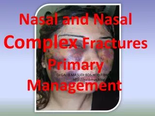
Nasal-Septal Fractures. Francis B. Quinn, M.D. Herve’ J. LeBoeuf, M.D. Anatomy. Bones - Frontal process of maxilla, nasal spine of frontal bone Paired nasal bones Vomer Perpendicular plate of the ethmoid. Anatomy (cont.). Cartilage- Lower lateral cartilage

E N D
Nasal-Septal Fractures Francis B. Quinn, M.D. Herve’ J. LeBoeuf, M.D.
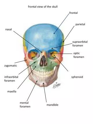
Anatomy Bones - • Frontal process of maxilla, nasal spine of frontal bone • Paired nasal bones • Vomer • Perpendicular plate of the ethmoid
Anatomy (cont.) Cartilage- • Lower lateral cartilage • Upper lateral (Alar) cartilage • Septal cartilage • Sesamoid cartilages
Pathogenesis Variables- • The patient’s age (tissue flexibility) • The amount of force applied • The direction of the force • The nature of the striking object
Frontal Impact Plane I- • Fracture of nasal tip • Small dorsal hump with supertip depression Plane II- • High fracture of nasal bones • Dorsal depression • Septal buckling with flattened appearance of the nose
Frontal Impact (cont.) Plane III- • Fracture of nasal bones, frontal process and anterior nasal spine • Comminuted, lateralized • Marked nasal depression • Columellar retraction • Medial canthal relaxation with telecanthus
Lateral Impact Plane I- • Unilateral nasal bone depression • Elevation of contralateral nasal bone • Septal buckling • C or S shaped deformity of nasal dorsum
Lateral Impact (cont.) Plane II/III- • Fracture extension to frontal process • Marked displacement of septum and dorsum • Medial maxillary wall depression
Septal Fracture • Vertical with anterior fracture • Horizontal with posterior fracture • S and C shaped deformities with healing • Telescoping of segments prevents closed reduction
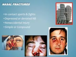
History • Force, direction of impact • Epistaxis • External deformity • Prior nasal injury, dysfunction • Pre-injury photographs
Exam • Nasal deviation • Mucosal or skin lacerations • Ecchymosis, hematoma • Lid edema, chemosis • Subconjunctival hemorrhage • Telecanthus, CSF rhinorrhea
Exam (cont.) • Topical decongestion • Debridement of clots • Internal and external palpation • Exam of cartilaginous nose • Roentgenograms • Photographic documentation
Clinical Decisions Open versus closed reduction Closed Reduction- • Unilateral or bilateral fracture of the nasal bones • Fracture of the nasal-septal complex with nasal deviation less than one half the width of the nasal bridge.
Clinical Decisions (cont.) Open Reduction- • Extensive fracture-dislocation of the nasal bones and septum • Nasal pyramid deviation exceeding one half the width of the nasal bridge • Fracture-dislocation of the caudal septum • Open septal fractures • Persistent deformity after closed reduction
Clinical Decisions (cont.) Local versus general anesthesia Timing of reduction- • < 3-6 hours- immediate reduction • < 2-3 weeks- closed reduction • > 3 weeks- delayed 3-6 months
Anesthesia • 4% cocaine • Epinephrine soaked pledgets • IV or oral sedation • EMLA cream - time consuming • General anesthesia
Instruments • Asch/Walsham forceps • Large Kelly clamps • Elevators- Boies/Ballinger • Various intranasal specula • Headlight
Reduction • Elevate fragment with anterolateral force • Completion of the fracture • External digital molding • Reduction of septum is critical • Asch/Walsham forceps to elevate fracture and reduce septum
Trouble Shooting • Overriding cartilage fragments • Post reduction instability • C-shaped septal fracture • Converting to an open reduction
Post-Op • Silastic splints • Intranasal placement of packing • External splint application • Packing out 2-3 days, silastic-10 days • External splint off when fracture stable
Subacute Open Reduction • Hemitransfixion, lateral intercartilaginous incisions • Elevation of dorsal skin and periosteum • Exposure of cartilage segments • Reduction of cartilage- scoring, suture • Maxillary crest involvement- “trapdoor”
Complicated Fractures • “Open sky” approach • Use preexisting lacerations when possible • Depressed comminuted fractures- wires versus miniplates • Wound closure • Prophylactic antibiotics
Delayed Repair • Complicated due to scarring, fibrosis • Common problems: Dorsal hump, C/S shaped septum, saddle deformities, septal displacement, fallen or deviated tip • Common solutions: Excision of hump, cartilage grafting, calvarial grafts, osteotomies
Children • Physical differences- projection, cartilage: bone, growth centers • Small fracture--- obstruction with age • Edema, anxiety tend to obscure fracture • Operative intervention- cosmesis, obstruction • Digital compression • Neonatal fracture-dislocation
Early Complications • Septal hematoma • Infections- antibiotic prophylaxis • Epistaxis- cautery, packing, ligation • CSF Rhinorrhea • Emphysema of the face, neck
Late Complications • Organization of hematomas- airway obstruction • Synechia- divide if symptomatic • Obstruction of the nasal vestibule • Residual osteitis • Malunion • Naso-facial disproportion