Download
1 / 20
230 likes | 607 Views
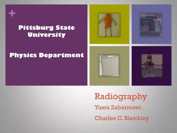
Pittsburg State University Physics Department. Radiography. Yusra Zabarmawi Charles C. Blatchley. Introduction. In this research, I design radiography through the use of a radioactive source. I use an animal phantom by using water instead of tissue and chicken bones .

E N D
Pittsburg State University • Physics Department Radiography YusraZabarmawi Charles C. Blatchley
Introduction • In this research, I design radiography through the use of a radioactive source. • I use an animal phantom by using water instead of tissue and chicken bones . • My detector is an autoradiography film and polaroid film.
Outline • What is radiography • Source of radiation • Geometry of shadow radiograph • My Researches Method and Materials Result • Conclusion
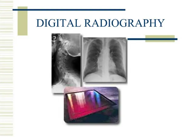
What is Radiography • Radiology is an area of medicine that utilizes imaging technology in order to diagnose and medicate illnesses. • It utilizes ionizing activating electromagnetic radiation
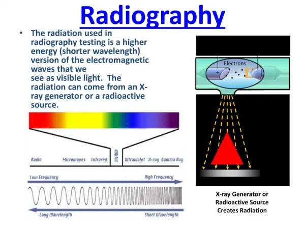
Radiation Source • Two of the most commonly used sources of radiation in radiography are x-ray generators and gamma ray sources. • X ray generators usually used on Medical Imaging . Gamma Ray
Geometry of shadow radiograph Source Object Detector
Detectors Radiographic film Flat Panel Detectors
“Gummite” is a generic term for a mixture of uranium-bearing minerals, usually consisting of Kasolite, Curite, Soddyite,Uranophaneand several others. • The exposure rate of Gummite is 2.4 mR/h at the surface.
Uranium Nitrate The exposure rate is 2.1 mR/h at the surface
Animal Phantom to represent the tissue I used a water inside the balloon And I use a chicken bones.
Polaroid film 4x5 Land film, Type 52, black and white. Autoradiography film by Carestream X-OMT LS film, size 13x18 cm .
2.0 5.9 inches 11 DAYS
11 DAYS 2.8 inches 2.0
11 DAYS 2.8 inches 2.0
10 DAYS 2.8 inches 1.0
Conclusion • In conclusion, radiography is a study that requires individual commitment for any scholar to be able to create it. • Radiography has offered positive contribution towards the development of human life especially in health. • A lot of research and invention should be practiced to perfect radiography.
References • Beutel, J., Harold, L., Kundel, R. & Metter, V. (Eds.). (2000). Handbook of medical imaging: Physics and Psychophysics (Vol. 1). Bellingham, WA: SPIE. • Clarke, C. &Dux, A. (2011). Chest x-rays for medical students. Hoboken, NJ: Wiley-Blackwell. • Dendy, P., & Heaton, B. (2012). Physics for diagnostic radiology (3rd ed.). Boca Raton, Fl: CRC Press. • Ritman, E. (2006). Medical x-ray imaging, current status and some future challenges. Department of Physiology & Biomedical Engineering. Retrieved from http:// www.icdd.com/resources/axa/vol49/v49_01.pdf