Download
1 / 35
510 likes | 1.86k Views
Radiography. Introduction. Objectives. To describe Properties of x-rays Production of x-rays Formation of radiographic image Components of an x-ray room The projections The radiographic technique. Kind of electromagnetic radiation Means of propagation of energy. What are x-rays?.

E N D
Radiography Introduction
Objectives To describe • Properties of x-rays • Production of x-rays • Formation of radiographic image • Components of an x-ray room • The projections • The radiographic technique
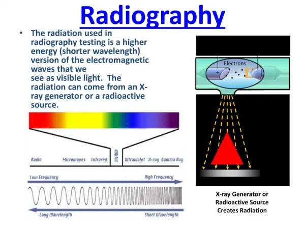
Kind of electromagnetic radiation Means of propagation of energy What are x-rays?
Nature of x-rays • Two theories to describe the nature 1. Wave theory (Propagate as waves) C = f λ ; Speed = frequency x wavelength 2. Corpuscular theory (Involved in interactions as particles) E = h f ; Photon Energy = Plank’s constant x Frequency
Properties/Characteristics of EM Radiation • Common properties • Properties Specific to x-rays
Characteristics of X-Rays • X-rays are invisible. • X-rays are electrically neutral. They have neither a positive nor a negative charge. • They cannot be accelerated or made to change direction by a magnet or electrical field. • X-rays have no mass. • X-rays travel at the speed of light in a vacuum. • X-rays cannot be optically focused. • X-rays form a polyenergetic or heterogenous beam.
The x-ray beam used in diagnostic radiography comprises many photons that have many different energies. • X-rays travel in straight lines. • X-rays can cause some substances to fluoresce. • X-rays cause chemical changes to occur in radiographic and photographic film. • X-rays can be absorbed or scattered by tissues in the human body. • X-rays can produce secondary radiation. • X-rays can cause chemical and biologic damage to living tissue.
Rectilinear propagation Penetration ability Differential absorption Fluorescent effect Photographic effect Ionization of medium Biological effects Prediction of the path Basis of radiography Tissue differentiation Intensification of image & fluoroscopy Image recording on photographic emulsions (films) Detection & measurement of radiation Radiotherapy & Need for radiation protection Properties & uses of x-rays
Production of X-rays X-rays are produced in an X-Ray tube by two processes: • Deceleration of fast moving electrons either by stopping, reducing speed or by changing its direction • The transfer of an electron between two inner orbits of an atom
Tap and sink • Hand gel • Detergent wipes • Soap and towel dispenser • Vomit bowels • Latex gloves / non latex gloves for those with allergies • Equipment needed for IVPs • General waste bin • Sharps bin • Emergency resuscitation mask/oxygen connectors • Oxygen outlet • Suction outlet
X-ray table • Overhead x-ray tube • Lead gowns • Moveable seat for patient positioning • Long leg x-ray film holder • Wall bucky • Arm extension for vertical bucky • External film holder • Contrast warmer • Filters for x-ray tube • Positioning Aids • Lead shielding
Principle of Image formation • X-ray beam of uniform intensity falls on object consists of structures with different absorption properties. • It attenuates in different amounts by different structures • The transmitted beam consists of different intensities – called the ‘aerial image’ • The aerial image falls on the image receptor, usually on a cassette containing x-ray film & intensifying screens
X-ray tube Plot of incident x-ray beam intensity Object Plot of transmitted x-ray beam intensity Invisible x-ray image
Invisible x-ray image kV mA Sec FFD E Supporting tissue (m) B B1 T3 B2 T1 T2 Air Invisible X-ray image consists of different x-ray intensities E B1 E B2 ET1 EM EM ET2 ET3 EA
Focal spot X-ray beam Cross section Plot of x-ray intensities
Radiographic projections • AP • PA • Lateral • Obliques:- RAO, LAO, RPO, LPO • Decubitus (patient lying down and the x-ray beam horizontal)
Description of Techniques • Position of patient • Position of body part • Position of cassette related to body part • Direction and centre of x-ray beam • Breathing status • Exposure factors • Radiographic appearance - criteria
Conclusion • The accuracy of diagnosis depends on the quality of the Image. • The radiographic image should have, • Optimum contrast • High resolution • Minimum unsharpness • Less noise • No artefacts • High definition
END Thank you