Download
1 / 41
410 likes | 488 Views
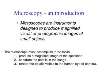
Microscopy - an introduction. Microscopes are instruments designed to produce magnified visual or photographic images of small objects. The microscope must accomplish three tasks 1. Magnify the specimen separate the details in the image,

E N D
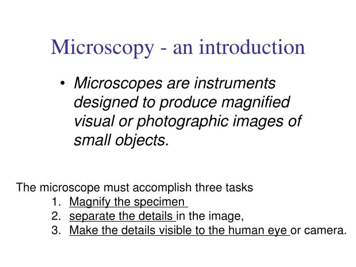
Microscopy - an introduction • Microscopes are instruments designed to produce magnified visual or photographic images of small objects. • The microscope must accomplish three tasks • 1. Magnify the specimen • separate the details in the image, • Make the details visible to the human eye or camera.
Microscopes increase resolution - minimumdistance two points can be apart and still be distinguished as two separate points
Microscope One or more lenses that make an enlarged image of an object.
Simple Microscope • Light passes through only 1 lens. • Example: magnifying glass
Compound Microscope • Lets light pass through an object and then through two or more lenses.
Stereoscopic Microscope • Gives a three dimensional view of an object. (Examples: insects and leaves) • Used for dissections
Electron microscopes – use a beam of electrons instead of a beam of light to magnify the image
Electron Microscopes • can achieve 3D images using electrons
The Scanning Electron Microscope • produces a 3-dimensional image of specimen’s surface features spider head of a butterfly
Scanning electron microscopy (SEM) • Types of specimens: • -Whole organisms • -Natural tissue surfaces • -Exposed tissue structure A flea magnified 50 000 X What is this?
Scanning Electron Microscope
Transmission electron microscopy (TEM). • Allows the observation of molecules within cells • Allows the magnification of objects in the order of 100, 000’s.
Longitudinal section of cilium Cross section of cilium 1 µm Transmissionelectronmicroscope (TEM) • Provides for detailed study of the internalstructure of cells • a beam of electrons is transmitted through the specimen for a 2D view Figure 6.4 (b) cilia on rabbit lungs
Transmission electron microscope Chloroplast from a tobacco leaf H1N1 virus
Confocal Laser Scanning Microscope (CLSM) • laser beam used to illuminate spots on specimen • computer compiles images created from each point to generate a 3-D image • used on specimens that are too thick for a light microscope
A, B, C pollen grains: Scanning electron microscope D pollen grains: Confocal Laser Scanning Microscope E pollen grains: Transmission electron microscope F pollen grains: Light microscope G Mixed pollen grains (bright field light microscope, stained) H pollen grains confocal laser scanning microscope
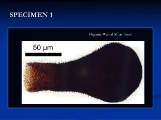
Look at the following micrographs (a picture made by a microscope) and try to determine what the object is!
What is the difference between a… VIRUS and CELL? E.coli bacterial cells
VIRUS BACTERIA • can’t live on its own- must - can exist on its own live inside another cell • much smaller (20 – 400nm) - larger (1000 nm = 1μm) • none are beneficial - some can be beneficial (bacteria in gut) • no cell wall, only a protein - outer cell wall coat - cannot be killed by antibiotics - are killed by antibiotics