Download
1 / 55
550 likes | 665 Views
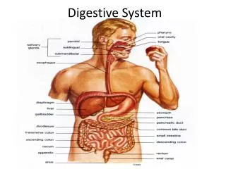
Digestive System. Digestion . Mechanical: breaks large pieces into smaller ones without altering their chemical composition. Chemical: breaks food into simpler chemicals. Alimentary Canal. Mouth Pharynx Esophagus Stomach Small Intestine Large Intestine Rectum Anus.

E N D
Digestion Mechanical: breaks large pieces into smaller ones without altering their chemical composition. Chemical: breaks food into simpler chemicals.
Alimentary Canal • Mouth • Pharynx • Esophagus • Stomach • Small Intestine • Large Intestine • Rectum • Anus
8 meters from mouth to anus. MOVEMENTS OF THE TUBE Peristalsis: propelling movements include a wavelike motion. - Ring of contraction appears in the wall of the tube, at the same time, the muscular wall just ahead of the ring relaxes.
The Oral Cavity Accessory organs • Tongue -frenulum, membranous folds that hold the tongue to the floor of the mouth. -papillae, projections, provide friction, taste buds. • Teeth • Salivary glands
Teeth • Incisors • Cuspid • Bicuspids (premolars) • molars
Enamel: covers the crown, calcium salts, hardest substance in the body. Dentin: substance like bone, harder, surrounds the tooth’s central cavity (pulp cavity). Pulp: blood vessels, nerves, Rootcanal: blood vessels and nerves go through. Cementum: bonelike material encloses the root Periodontalligament: surrounds cementum, attaches tooth to jaw.
Salivary Glands • Parotid • Submandibular • Sublingual Saliva is comprised of water (99.4%) with an assortment of ions, buffer, metabolites, and enzymes. Collectively, the salivary glands produce 1-1.5L/day
Palate Palate: roof of the oral cavity, hard anterior part (hard palate). Uvula: coneshaped projection from the soft palate. During swallowing the uvula is drawn upward. This action closes the opening between the nasal cavity and the pharynx, preventing food from entering the nasal cavity.
Palatinetonsils: lymphatic tissue, help protect the body against infection. Pharyngealtonsils (adenoids): posterior wall of the pharynx, above the border of the soft palate. If they enlarge and block the passage between the pharynx and the nasal cavity, they may be removed.
1. Esophagusperistalsis pushes food to stomach. • Peristalsis wavelike motion that propels food. 2. Stomachsecretes acid and enzymes. Mixes food with secretions to begin enzymatic digestion of proteins.
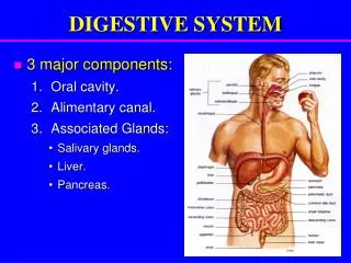
Digestive System Structures Digestive tract Accessory organs • Teeth • Tongue • Salivary glands • Liver • Gall bladder • pancreas Oral cavity Pharynx Esophagus Stomach Small intestine Large intestine
Small Intestine mixes food with bile and pancreatic juice. Final enzymatic breakdown of food molecules: main site of nutrient absorption. • Liver produces bile, which emulsifies fat. • Gallbladder stores bile and introduces it into small intestine. • Pancreas produces and secretes pancreatic juice, containing digestive enzymes and bicarbonate ions, into sm. Intestine. • Accessory organs are organs that are not a direct route of the digestive system.
Large intestine absorbs water and electrolytes to form feces. • Rectum regulates elimination of feces. • Anus eliminates feces. • The digestive system is a tube, open at both ends, that has a surface area of 186 square meters. • The main function is to supply body with nutrients.
Alimentary Canal • Mucosa or mucous membrane the inner most layer. Contains glands that secrete mucus and digestive enzymes. Carries on secretion and absorption. Also, has projections that increase the surface area. Lumen=passageway. • Submucosa contains loose connective tissue, glands, blood vessels, lymphatic vessels, and nerves. Carry away absorbed materials.
Muscular layer produces the movements. • Serosa or serous layer the outer covering of the digestive tube. Secretes fluid that lubricates the tube’s outer surface so organs slide freely against one another.
The Pharynx 3 Divisions: Nasopharynx Oropharynx Laryngopharynx The pharynx is a muscular structure with as epithelial lining that functions in both respiration and digestion. Pharyngeal muscles cooperate with the oral cavity and esophagus to initiate the swallowing process
Three Phases of Swallowing Buccal (Oral)Phase Pharyngeal Phase Esophageal phase
During swallowing, muscles draw the soft palate and uvula upward to separate the oral and nasal cavities. • Salvia cleanse the mouth and teeth, dissolve chemicals necessary to tasting food, and help in the formation of food bolus.
Tonsils • Palatine are lymphatic tissue in the back of the mouth, on either side of the tongue and closely associated with the palate. Help to fight infection. • Pharyngeal or adenoids are on the posterior wall of the pharynx. They can be removed. • Tonsillectomy tonsil are surgically removed.
Teeth • 4 types of teeth • Incisors (Front 4 teeth) bite off pieces of food. • Cuspid grasp and tear food. • Bicuspids or premolars grind food. • Molars grind food.
The Stomach *The mixing of ingested substances with the gastric juices (secreted by gastric glands) produces chyme. • Landmarks: • Lesser curvature • Greater curvature • Functions: • Bulk storage of ingested food • Mechanical breakdown of ingested food • Chemical digestion of ingested food via the disruption of chemical bonds by acids and enzymes
Stomach • The stomach is divided into four regions. • Cardiac • Fundic is the area that acts as a temporary storage area for ingested food. • Body • Pyloric • Fatty food stay in the stomach the longest. Pyloric sphincter (empties into small intestines)
Gastric Gland Secretions • Mucous cells: secrete alkaline substance that protects stomach. • Parietal cells: secrete HCL • chief cells: secrete digestive enzymes.(pepsinogen) Makes gastric juice
Gastric Secretions • Gastric juice • Hydrochloric acid • Pepsin begins to digest protein (pepsinogen meets HCL and forms pepsin, active) • Chyme is a semifluid paste of food particles and gastric juice.
Intrinsicfactor: parietal cells secrete. Helps the small intestine absorb vitamin B12. Vagus nerves stimulate the release of Ach from nerve endings. Simulates gastric glands to secrete abundant gastric juice. Occurs when: tastes, smells, or even sees appetizing food, or when food enters stomach. Gastrin: hormone that increases the secretion of gastric juice.
Time Spent in organs • Mouth a few minutes • Esophagus a few minutes • Stomach 4 hours • Small Intestine 12 hours • Large Intestine 5 hours • Total time in digestive system 21 hours.
The Small Intestine Duodenum – “mixing bowl” receives chyme from stomach and digestive enzymes from pancreas and liver Jejunum – bulk of chemical and nutrient absorption Ileum– controls flow of material into the large intestine via Pyloric Sphincter
Absorptive structures • Villi • These structures function to increase the surface area of the inner intestinal wall thereby facilitating absorption.
The Large Intestine • Divisions: • Cecum • Colon • Rectum • Functions: • Re-absorption of water and electrolytes • Compaction of intestinal contents into feces • The absorption of vitamins
Rectum and Anus Internal anal sphincter – involuntary smooth muscle. External anal sphincter – voluntary skeletal muscle whose relaxation allows for defecation
Gall Bladder The gallbladder is a sac located under the liver. It stores and concentrates the bile produced in the liver. Bile is released from the gallbladder in response to food, especially fats, in the upper small intestine.
15.7 Pancreas • Pancreatic juice • Pancreatic acinar: cells that produce pancreatic juice. • Pancreatic duct: connects with duodenum.
Pancreatic Juice • Digests: carbs, fats, nucleic acids, and proteins. • Pancreaticamylase: carb digesting enzyme. • Pancreaticlipase: fat-digesting enzyme. • Nucleases: enzymes that break down nucleic acid molecules into nucleotides.
Regulation Trypsin, chymotrypsin, carboxypeptidase: protein-splitting enzymes. Secretin: hormone that stimulates secretion of pancreatic juice, occurs when chyme enters the duodenum. Cholecystokinin: travel to pancreas and stimulates pancreatic juice secretion.
15.8 Liver • The liver is the largest organ in the body it is the heaviest organ in the body at around 3 pounds. • Divided into lobes: right lobe (larger) and left lobe (smaller), separated into many tiny hepatic lobules, liver’s functional units. • Centralvein: in a lobule, hapatic cells radiate outward from a central vein. • Hepaticsinusoid: vascular channels, separate platelike groups of cells from each other.
Hepatic portal vein: brings newly absorbed nutrients into the sinusoids and nourishes the hepatic cells. • Kupffer cells: phagocytic macrophages are found in the hepatic sinusoids. Remove bacteria and other foreign substances. • Bile is made in liver and converge to the common hepatic duct.
Functions • Maintain normal concentration of blood glucose levels. • Liver cells respond to insulin and glucagon, lower the blood glucose level. (glycogen). • Digests lipids. • Protein metabolism: makes plasma proteins, covert amino acis. • Stores: glycogen, iron, and vitamins A, D, and B12.
Liver contains macrophages that help destroy damaged red blood cells. • Liver removes toxic substances such as alcohol from blood.
Bile • Yellowish green liquid secreted from hepatic cells. • Bile salts, bile pigments (bilirubin and biliverdin), cholesterol, and electrolytes. • Bile pigments breakdown products of hemoglobin from red blood cells and are normally excreted in the bile.
Gallbladder • Joins the common hepatic duct. • Cystic duct, right off gallbladder. • Stores bile between meals, reabsorbs wter to concentrate bile, releases bile into the small intestine. • Hepatic and cystic cuts join to form common bile duct. • Cholecystectomy: removal of gallbladder.
Bile salts break fat globules, action called emulsification.
Digestive enzymes • Salivary glands make chemicals that digest only carbohydrates. • Pancreas makes chemicals that digest fat, protein, and carbohydrates. • Liver makes chemicals that digest only fat. • Stomach makes chemicals that digest only protein. • Small intestine makes chemicals that digest protein and carbohydrates. • Large intestine makes no chemicals to digest food.
http://highered.mcgraw-hill.com/sites/0072351187/student_view0/chapter15/chapter_quiz.htmlhttp://highered.mcgraw-hill.com/sites/0072351187/student_view0/chapter15/chapter_quiz.html
15.9 Small Intestine • Receives secretions from the pancreas and liver. • Completes digestion of the nutrients in chyme. • Absorbs the products of digestion. • Transports residue to large intestines.
Parts of Small Intestine • Duodenum – C shaped • Jejunum • Ileum • Mesentery: double-layered fold of periotoneal membrane, supports blood vessels, nerves, and lymphatic vessels that supply the intestinal wall.
Structure • Intestinalvilli: projections of mucous membrane, increase the surface area for absorption.