Download

1 / 12
150 likes | 576 Views
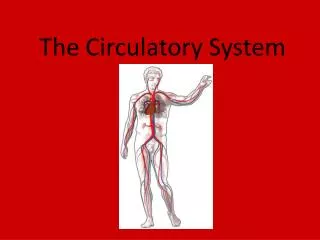
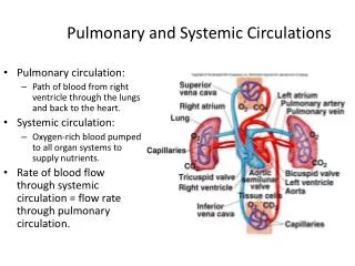
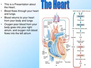
The Pulmonary Circuit (blood flow between heart and lungs). Follow a drop of blood through the path seen on the next slide. Understand that blood flow between the aorta and the vena cavas constitutes the Systemic Circuit .

E N D
The Pulmonary Circuit(blood flow between heart and lungs) • Follow a drop of blood through the path seen on the next slide. Understand that blood flow between the aorta and the vena cavas constitutes the Systemic Circuit. • PSV: pulmonary semilunar valve. ASV: aortic semilunar valve. Remember not to abbreviate anything on tests in this class except M, N, A, V, T, L. • The Tricuspid valve is AKA the Right Atrioventricular Valve. The Bicuspid valve is AKA the Mitral or Left Atrioventricular Valve. • Note that the entire right side of the heart pumps low-oxygen blood, while the entire left side of the heart pumps high-oxygen blood.
The Pulmonary Circuit SVC/IVC Right Atrium Tricuspid valve Right Ventricle PSV Pulmonary AA Capillaries of lungs Pulmonary VV Left Atrium Bicuspid Valve Left Ventricle ASV Aorta/AA
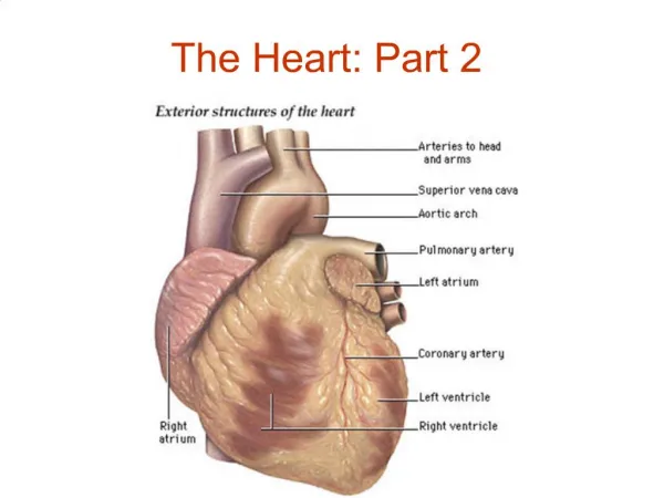
Chambers of the heart • The atria receive blood from either the vena cavas (right) or the pulmonary veins (left) and pump blood through the atrioventricular valves to the ventricles. Typically, only the right atrium has the myocardial features inside known as pectinate muscles. These criss-crossed muscle bands end on a smooth band inside called the crista terminalis. Also better seen in the right atrium is a depression on the atrial septum called the fossa ovalis. This feature is discussed later in fetal circulation. • The ventricles are far more muscular than the atria, with the left ventricle having the thickest myocardium of all chambers. On the walls of both ventricles are muscular features called trabeculae carnae. Other myocardial features are the papillary muscles, which connect to the Atrioventricular (A/V) valve cusps via chordae tendineae. This arrangement of structures allows the A/V valves to close tightly when the ventricles contract.
Chambers of the heart • Unique to the Right Ventricle is the moderator band – a stabilizing band of myocardium between the largest papillary muscle and the interventricular septum. • Each ventricle, when contracted muscularly, causes closure of its A/V valve and opening of a semilunar valve. The upper part of the right ventricle narrows to a cone-shaped portion called the conus arteriosus, which is visible just below the pulmonary semilunar valve. At the base of the aorta, within the left ventricle, is the aortic semilunar valve. • This is all the same anatomy you’ve been learning in lab, now know it for lecture.
Internal thoracic structures HEART RIGHT ATRIUM
Internal thoracic structures HEART RIGHT VENTRICLE
Internal thoracic structures HEART LEFT VENTRICLE
The Conducting System of the Heart(innervation of the myocardium) • The Sinuatrial (SA) node is a ganglion at the base of the SVC and the Right atrium which produces impulses about 70 times per minute. It is innervated sympathetically by fibers from the sympathetic trunks and parasympathetically by the Vagus N. This is the heart’s natural pacemaker, which can even operate without any outside nerve stimulation at all. • Every time the Sinuatrial node ‘fires’ the atria contract muscularly, which stimulates another ganglion within the upper ventricular septum: the Atrioventricular (AV) Node. The atrioventricular node then sends an impulse down the atrioventricular bundle (aka Bundle of His) to nerve endings known as Purkinje Fibers, which are found in ventricular walls. By the time stimulation reaches Purkinje Fibers, the ventricles are fully contracting. • One complete cycle of atrial and ventricular contraction is called a Cardiac Cycle. With reference to the heart, contraction of chambers is called systole, while relaxation and filling is called Diastole. Thus, the peak of blood pressure occurs during ventricular systole (systolic) and the minimum pressure occurs during ventricular diastole (diastolic).
Sinuatrial node (SA node) Atrioventricular node (AV node) -Bundle of His in the interventricular septum -Purkinje fibers stimulate muscle of ventricles CONDUCTING SYSTEM OF THE HEART
Along with learning about fetal circulation, be sure to know all the coronary AA and cardiac VV you studied in lab for lecture as well.
Blood flow in the fetus In the human fetus (period between 3rd month and birth) the lungs are not being used for respiration and so do not require as much blood as adult lungs. Also, the mother is providing oxygenated blood to the developing child via the umbilical blood vessels. -Umbilical vein caries O2 blood to fetus >Mixes with low O2 blood in IVC >Blood enters the R atrium and then passes through the foramen ovale (later the fossa ovalis) to the L atrium to bypass the pulmonary circuit. >Some blood enters the R ventricle and is sent to the Pulmonary trunk. >Blood in the Pulmonary trunk is shunted to the Aorta though the Ductus Arteriosus (later becomes the ligamentum arteriosum). -The result is that there is a mixing of high and low O2 blood in the fetus, and the lungs only receive a small amount of blood.
Fetal circulation continued -The Foramen ovale and the Ductus Arteriosus close muscularly (like sphincters) at birth with the first breaths of the infant. -The Ductus Arteriosus will ‘fuse’ closed within about 3 months. -Fusion of the Foramen ovale takes about 1 year. Failure of either or both of these fetal openings to close results in “blue baby syndrome”. Problems with the closure of these openings account for about 8% of congenital heart defects. An opening in the fossa ovalis, for example, is called an Atrial Septal Defect (ASD) and can be seen in about 25% of the population. Small openings here tend to be asymptomatic.