Download
1 / 26
280 likes | 1.14k Views
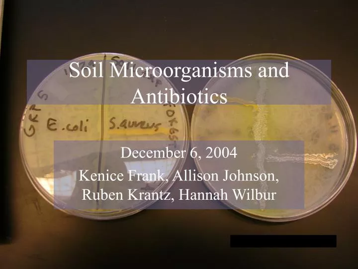
Soil Microorganisms and Antibiotics. December 6, 2004 Kenice Frank, Allison Johnson, Ruben Krantz, Hannah Wilbur. Habitats: Water films Need water for metabolic processes On surface of organic matter Need surface on which to grow In rhizosphere. Competition

E N D
Soil Microorganisms and Antibiotics December 6, 2004 Kenice Frank, Allison Johnson, Ruben Krantz, Hannah Wilbur
Habitats: Water films Need water for metabolic processes On surface of organic matter Need surface on which to grow In rhizosphere Competition Compete with surrounding bacteria and fungi Production of antibiotics by competing bacteria Soil bacteria Soil environments are host to a great number of bacterial species
Can be used as: Fungicide, etc for farming Antibiotics for humans and animals From Strepomyces species alone: 500 antibacterial products identified Antibiotics
Objectives • Identify through laboratory testing members of the soil communities • Isolate and cultivate antibacterial producing bacteria from soil • Observe and understand members of different communities of soil bacteria
Soil Experiment Methods and Materials
Methods and Materials • The first step of our experiment was to chose 3 different locations/environments & to obtain soil samples. We chose a river, marsh and forest. Marsh Forest River • We then used sterile tubes to obtain the sample
Methods and Materials • We added sterilized H2O to each soil sample and streaked 500 ml of each soil type onto 4 starch-casein agar for each soil type. • We incubated these plates at room temperature.
Methods and Materials Week 2 • We took 100 ml of forest sample and diluted it with 400 ml more of sterile H2O • We isolated 4 visually different colonies from each soil type and streaked each colony type onto individual starch casein agar -We did a smear of the entire forest plate and the entire river plate because there were no identifiable, separate colonies • Incubate at room temperature
Methods and Materials Week 3 • Got results from previous week’s plates & examined for any Streptomyces by looking for any areas of inhibition (clear areas surrounding colonies) • Using soil plate prepared for the general lab, we tried to isolate antibiotic producing organism again Agar plates showing areas of inhibition
Methods and Materials • We streaked the lab specimen and a sample from each soil plate from last week down the middle of a BAP (blood agar plate) and then used method for testing sensitivity to observe for any areas of inhibition or hemolysis • We then streaked S.aureus and E.coli on starch casin agar plates perpendicularly to a center streak of each isolated colony type without touching it Blood Agar Plate (BAP)
Methods and Materials • We performed Gram staining on -3 forest plates -2 river plates -4 marsh plates • Based on our results from the Gram staining, we decided that we would need to identify whether we had any bacilli or enterics by performing some tests 1.Bacillus- endospore staining, catalase test 2.Enterics- oxidase test, TSI test • Make decision about what colonies were growing on the plates
Soil Experiment RESULTS
Starch Casein Plates • 1st plating: cultures too thick • 2nd plating: individual colonies observed Gram Stain: Marsh A:Gram (+) & Gram (-) rods Marsh B: Gram (+) rods Marsh C: Gram (-) cocci River A: Gram (-) cocci in clusters River B: Gram (-) cocci in clusters Forest A: Gram (-) rods Forest B:Gram (+) ovals Forest C: Gram (-) cocci in clusters
Sensitivity/Inhibition Testing • No inhibition observed
Blood Agar Plate • Marsh B: total hemolysis • Forest A: total hemolysis
Forest A (Gram neg. rods): Oxidase positive Forest A: Catalase positive Marsh B: Catalase positive Oxidase Test Catalase Test
Triple Sugar Iron Test • Forest A: Red/Yellow (K/A) Glucose and 1 other sugar fermented
Endospore Staining • Red bacillus cells • No endospores observed
Soil Experiment Discussion
Discussion The first goal was to properly identify Streptomyacin, or other inhibiting agents produced by the bacteria in the soil that would supposedly combat against E. coli and S. aureus. These attempts failed, as both species of bacteria sustained growth.
Discussion The experiment was also intended to isolate certain colonies from three different environments: forest, marsh, and river. After single colonies of the first plating were isolated physically, a gram stain from the marsh indicated that bacillus and enteric species were present. Tests resulted negative for enteric bacteria and positive for bacillus, however the endospore stain resulted negatively.
What should have happened: After the second plating the colonies of E. coli and S. aureus should have showed suppressed growth to any inhibiting factors that the samples produced. Tests for Streptomyacin
What went wrong: The procedure for isolating the bacteria should have been done using selective and differential media in order to eliminate any other contamination in the culture. Other possibilities are that streptomyacin producing bacteria did not, in fact, reside at the chosen locations. Tests for Streptomyacin
Enteric bacteria are gram negative rods. They are usually Oxidase negative and Catalase positive. They are nitrate reducers, as they are commonly found in some soils. They are also known to ferment glucose. Bacillus species are gram positive rods. They are endospore forming and are Catalase positive. They are also hemolytic. Bacterial Identification
What went wrong: The enteric tests showed Oxidase positive which is not a characteristic of enteric bacteria. The tests for the Bacillus colonies were all correct, however the endospore stain did not show spore production. This could be because the bacteria was not in an environment that spore production was needed. The second plating also should have been done using selective and differential agar to remove any contaminations. The secong plating should have been inoculated from the same spot on the first petri dish. Other possibilities are that these could be mutants. Bacterial Identification
References • Fenchel T. 2001. Bacterial Ecology. In Encyclopedia of Life Sciences. www.els.net • Madigan, MT, Martinko M, Parker J. 2003. Filamentous, High GC, Gram-Positive Bacteria: Streptomyces and other Actinomycetes. In Brock Biology of Microoganisms. pp. 416-420. New Jersey: Pearson Education, Inc. • Davelos AL, Kinkel LL, Samac DA. 2004. Spatial Variation in Frequency and Intenstiy of Antibiotic Interactions among Streptomycetes from Prairie Soil. Applied and Environmental Microbiology 70(2); 1051-58.