Download
1 / 73
770 likes | 1.05k Views
The Autonomic Nervous System and the Adrenal Medulla. Prof. dr. Zoran Vali ć Department of Physiology University of Split School of Medicine. portion of the nervous system that controls most visceral functions of the body

E N D
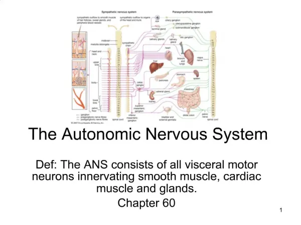
The Autonomic Nervous System and the Adrenal Medulla Prof. dr. ZoranValić Department of Physiology University of Split School of Medicine
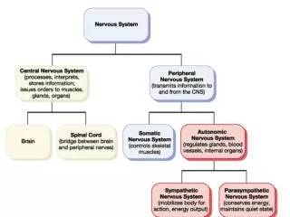
portion of the nervous system that controls most visceral functions of the body • arterial pressure, gastrointestinal motility, gastrointestinal secretion, urinary bladder emptying, sweating, body temperature • rapidity (seconds)andintensityof the change (100%) – most striking characteristics
General Organization of the ANS • centers located in: • spinal cord • brain stem • hypothalamus • + limbic cortex • operates through visceral reflexes (subconscious) • sympathetic and parasympathetic nervous system
Physiologic Anatomy of the Sympathetic Nervous System • two paravertebral sympathetic chains of ganglia • two prevertebral ganglia • celiac • hypogastric • postganglionic nerves • sympathetic nerve fibers originate in the spinal cord along with spinal nerves between cord segments T-1 and L-2
Preganglionic and Postganglionic Sympathetic Neurons • difference from skeletal motor nerves • cell body of each preganglionic neuron lies in the intermediolateral horn of the spinal cord • its fiber passes through an anterior root of the cord into the corresponding spinal nerve
after the spinal nerve leaves the spinal canal, the preganglionic sympathetic fibers leave the spinal nerve • pass through a white ramus into one of the ganglia of the sympathetic chain
course of the fibers can be following: • synapse with postganglionic sympathetic neurons in the ganglion that it enters • pass upward or downward in the chain and synapse in one of the other ganglia of the chain • pass for variable distances through the chain and then through one of the sympathetic nerves radiating outward from the chain, finally synapsing in a peripheral sympathetic ganglion • postganglionic fibers then travel to their destinations in the various organs
some of the postganglionic fibers pass back from sympathetic chain into spinal nerves through gray rami at all levels of the cord • these are all very small type C fibers, and they extend to all parts of the body by way of the skeletal nerves • they control: blood vessels, sweat glands, and piloerector muscles of the hairs • they make 8% of fibers in average nerve
Segmental Distribution • sympathetic pathways are not necessarily distributed to the same part of the body as the somatic spinal nerve fibers from the same segments: • T1: head • T2: neck • T3-T6: thorax • T7-T11: abdomen • T12-L2: legs
distribution is only approximate and overlaps greatly • it is determined partly by the locus in the embryo from which the organ originated
Adrenal Medullae • preganglionic sympathetic nerve fibers pass, without synapsing, from the intermediolateral horn cells of the spinal cord, through the sympathetic chains, then through the splanchnic nerves, and finally into the two adrenal medullae
these secretory cells embryologically are derived from nervous tissue and are actually themselves postganglionic neurons (they even have rudimentary nerve fibers) • the endings of these fibers that secrete the adrenal hormones epinephrine and norepinephrine
Physiologic Anatomy of the Parasympathetic Nervous System • parasympathetic fibers leave the CNS through cranial nerves III, VII, IX, and X, additionally by 2nd and 3rd sacral spinal nerves (sometimes 1st and 4th) • 75 percent of all parasympathetic nerve fibers are in the vagus nerves • vagus nerves supply: heart, lungs, esophagus, stomach, entire small intestine, proximal half of the colon, liver, gallbladder, pancreas, kidneys, and upper portions of the ureters
Preganglionic and Postganglionic Parasympathetic Neurons • preganglionic fibers pass uninterrupted all the way to the organ that is to be controlled (except in the case of a few cranial nerves) • postganglionic neurons are located in the wall of the organ • postganglionic fibers, a fraction of a millimeter to several centimeters in length, leave the neurons to innervate the tissues of the organ
Cholinergic and Adrenergic Fibers • cholinergic fibers – secrete acetylcholine • adrenergic fibers – secrete norepinephrine (noradrenalin)
all preganglionic neurons (fibers) are cholinergic (sympathetic and parasympathetic) • all or almost all of the postganglionic neurons (fibers) of the parasympathetic system are also cholinergic • most of the postganglionic sympathetic neurons (fibers) are adrenergic
exemption: postganglionic sympathetic nerve fibers to the sweat glands, to the piloerector muscles of the hairs, and to a very few blood vessels are cholinergic
acetylcholine – parasympathetic neurotransmitter • norepinephrine – sympathetic neurotransmitter
Secretion ofACh and NE • few parasympathetic nerve endings are similar to, but much smaller, than those of the skeletal neuromuscular junction • majority of fibers merely touch the effector cells of the organs that they innervate • varicosities – bulbous enlargements – ACh and NE, mitochondria • action potential entrance of Ca secretion from terminals or varicosities
ACh • synthesized in the terminal endings and varicosities • stored in vesicles • acetyl-CoA + choline acetylcholine • acetylcholinesterase from local connective tissue splits it to acetate ion and choline • choline secreted is then transported back into the terminal nerve ending (reuptake)
NE • synthesis in nerve endings, but is completed inside the secretory vesicles • tyrosine (hydroxylation) DOPA (decarboxylation) dopamine (transport into vesicles, hydroxylation) NE (in AM, 80%, methylation) epinephrine • removal: • reuptake (50-80%) • diffusion into surrounding body fluids (most of the remaining NE) • enzymatic degradation (MAO, COMT, small amounts)
NE secreted directly into a tissue remains active for only a few seconds • NE and epinephrine secreted into the blood by the AM remain active until they diffuse into some tissue (COMT, liver) • NE and epinephrine remain active for 10 to 30 seconds; but their activity declines to extinction over 1 to several minutes
Receptors • ACh and NE must first bind with specific receptors on the effector cells • receptor is on the outside of the cell membrane • binding of transmitter substance causes a conformational change in the structure of the protein molecule • change in cell membrane permeability • activating or inactivating an enzyme
Change in Membrane Permeability • opening or closing an ion channel • most frequently sodium and/or calcium ion channels • entrance of ions usually depolarizes the cell membrane and excites the cell • exit of K ions from the cell leads to hyperpolarization (inhibition) of the cell
Altering Intracellular "Second Messenger" Enzymes • binding NE adenylylcyclase cAMP • exact effect depends on the chemical machinery of the effector cell
Acetylcholine Receptors • nicotinic receptors • activated by nicotine • muscarinic receptors • activated by muscarine (a poison from toadstools) • AChactivates both nicotinic and muscarinic receptors
1. Nicotinic receptors • found at the synapses between the preganglionic and postganglionic neurons of both the sympathetic and parasympathetic systems • also present at many nonautonomic nerve endings; at the neuromuscular junctions in skeletal muscle
ionotropic receptor (directly connected with the ion channel, does not utilize second messengers) • two subclasses: • nicotinic receptor of the muscle type • nicotinic receptor of the neuronal type
2. Muscarinic receptors • found on all effector cells that are stimulated by the postganglionic cholinergic neurons • either the parasympathetic nervous system or the sympathetic system
metabotropic receptor (receptor coupled with G protein) • five subclasses (M1-M5)
Adrenergic Receptors • α-receptors • in turn divided into α1- i α2-receptors • β-receptors • in turn divided into β1-, β2- i β3-receptors • NE excites mainly α-receptors, β-receptors to a lesser extent • epinephrine excites both types of receptors approximately equally
metabotropicreceptor (receptor coupled with G protein) • under the influence of catecholamins • isopropyl norepinephrine (isoprenaline or isoproterenol) acts on β-receptor
Excitatory and Inhibitory Actions • excitatory effects in some organs but inhibitory effects in others – there is no generalization,one must learn all the separate functions of these two nervous systems on each organ • when sympathetic stimulation excites a particular organ, parasympathetic stimulation sometimes inhibits it
most organs are dominantly controlled by one or the other of the two systems
Function of the Adrenal Medullae • stimulation of the sympathetic nerves to the AM causes large quantities of epinephrine (80%) and NE (20%) to be released, although proportions can differ • almost the same effects as direct sympathetic stimulation, except that the effects last 5 to 10 times as long (2-4 min)
epinephrine has a greater effect on cardiac stimulation, less vasoconstrictive effect (especially in muscle, represent a major segment of the vessels of the body) • NE greatly increases the total peripheral resistance and elevates arterial pressure; epinephrine raises the arterial pressure to a lesser extent but increases the cardiac output more
epinephrine has 5 to 10 times as great a metabolic effect as NE
Value of the Adrenal Medullae to the Function of the SNS • stimulation of organs in two ways: directly by the sympathetic nerves and indirectly by the adrenal medullary hormones • two means of stimulation support each other, sometimes substitution for the other – safety factor