Download
1 / 24
240 likes | 308 Views
Develop knowledge of microscopy and microbial observation techniques. Learn to discern and identify different bacteria types. Practice aseptic techniques and isolation methods for pure cultures. Understand bacterial contamination and growth media.

E N D
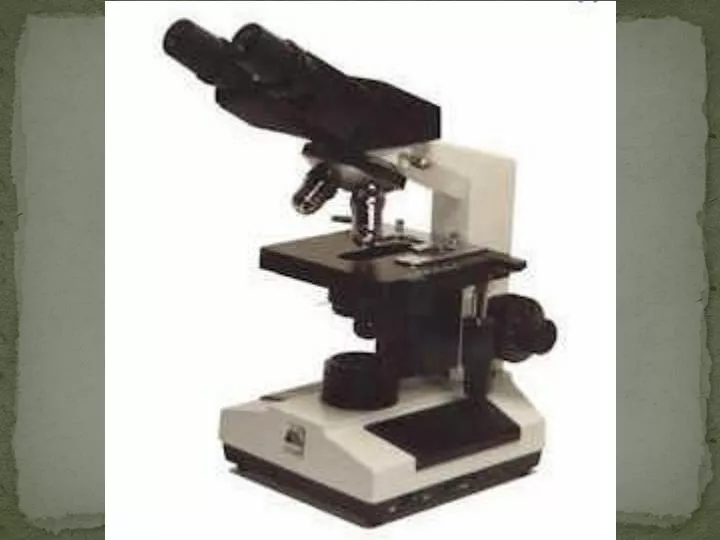
Exercise 2: 2A: Microscopy
Announcements • Post Lab 2 is assigned today and due by the time your lab meets next. • Pre Lab 3 will be available on Wednesday at 5 PM and is also due by the time your lab meets next. • LNA Bacteria is assigned today, and due by the time your lab meets next*. • Pre-Lab Write for LNA 3 is due within the first 5 minutes of lab next week.
Goals • Develop working knowledge of a brightfield light microscope • Discern between different types of microscopy • Practice techniques: objectives, oil, wet mount, measurements
Practice • Practice with the objectives, focusing, and positioning using the prepared slides. • Use your lab manual as a reference if you are having trouble pages 29-30.
Observation of Bacteria • Sphere • cocci • Rod • bacilli • Spiral • Spirochete and spirilla
Exercise 2: 2B: Bacteria
Goals • Become familiar with the scientific process by generating hypotheses, making predictions and designing experiments • Determine potential sources of microbial contamination in the laboratory • Obtain a pure culture of bacteria by streaking for isolated colonies on solid media
Bacteria • Prokaryotes: lack a nuclear membrane • Small, single cell organisms • Exist in huge numbers in small amounts of material • Found almost everywhere
Contamination • Look for contamination by testing for growth on bacterial medium.
Pure Cultures • A population of cells, all of which are descended from a single cell.
Growth Media • Liquid or Solid • Agar • Non-toxic • Remains solid at high temperatures • Not used as a nutrient • Defined or Complex
Sterilization • To kill all living organisms • Autoclaving • Baking • Alcohol • Flaming • Filtration
Aseptic Technique • Inoculating Loop • Serological Pipettes • Microliter Pipettes
Colony • Cluster of cells visible to the naked eye • Facilitating: • Isolation of pure cultures • Enumeration of cell concentration in liquid suspensions • ID of bacterial species based upon the appearance of the colonies
Spreading Plates • A diluted suspension is pipetted directly onto the surface of an agar plate and spread across the surface using a sterile glass spreader.
Determining the number of viable bacteria • The number of colonies on a plate is assumed to be equal to the number of viable cells which were spread on the plate • Limiting Factors • No more than 0.2 ml of cell suspension should be spread on the plate • Resulting in 30 to 300 cells spread on the plate
Determining Cell Mass by Measuring Turbidity • The number of cells per ml is directly proportional to the mass of cells per ml • Using Spectrophotometers to measure Optical Density (OD) • Light is lost as it passes through the suspension because it is scattered and absorbed by the cells. • Data can be used to construct a standard curve.
Bacterial Stains • Positive Stains: cells pick up color • Negative Stains: background color, cells appear white
Procedure Part I • Labeling plates • Label the bottom of the agar plate • Write on the periphery of the plate • Write your name, section, date, and location • Use the sterile swab to collect your contaminant and put it on the agar plate • Incubate plates for 48 hours at 37ºC
Procedure Part II • Isolate Pure Cultures • Using Aseptic Technique • E. coli • Use Streak Techniques
First LNA: Due Next Lab • You should come to lab next with with your lab notebook assignment. • There will be enough time at the beginning of next lab to observe your plates and finish the results and conclusions. • Also remember to have your Pre-Lab write-up (purpose, procedure, data table) for Exercise 3: Enzymes at the beginning of class.