Download
1 / 50
690 likes | 2.08k Views
ANDALIB.ALI MD Alzahra hospital. PEDIATRIC SUPRACONDYLAR HUMERUS FRACTURE. Supracondylar Humerus Fractures. Most common fracture around the elbow in children (60 percent of elbow fractures) 95 percent are extension type injuries, which produces posterior displacement of the distal fragment

E N D
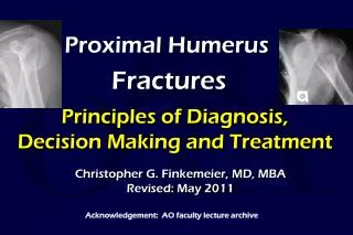
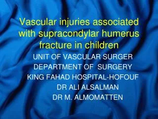
ANDALIB.ALI MD Alzahra hospital PEDIATRIC SUPRACONDYLAR HUMERUS FRACTURE
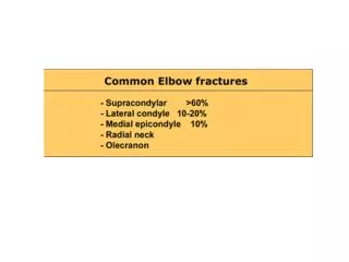
Supracondylar Humerus Fractures • Most common fracture around the elbow in children (60 percent of elbow fractures) • 95 percent are extension type injuries, which produces posterior displacement of the distal fragment • Occurs from a fall on an outstretched hand • Ligamentous laxity and hyperextension of the elbow are important mechanical factors • May be associated with a distal radius or forearm fracture
SupracondylarHumerus Fractures:Classification • Gartland (1959) • Type 1 non-displaced • Type 2 Angulated/displaced fracture with intact posterior cortex • Type 3 Complete displacement, with no contact between fragments
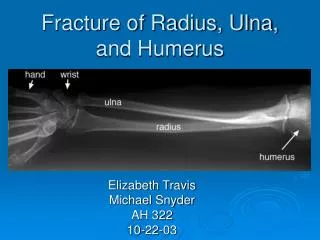
Elbow Fractures in Children:Radiograph Anatomy/Landmarks • Anterior Humeral Line: This is drawn along the anterior humeral cortex. It should pass through the middle of the capitellum.
30 Elbow Fractures in Children:Radiograph Anatomy/Landmarks • The capitellum is angulated anteriorly about 30 degrees. • The appearance of the distal humerus is similar to a hockey stick.
Elbow Fractures in Children:Radiograph Anatomy/Landmarks • The physis of the capitellum is usually wider posteriorly, compared to the anterior portion of the physis Wider
Elbow Fractures in Children:Radiograph Anatomy/Landmarks • Radiocapitellar line – should intersect the capitellum • Make it a habit to evaluate this line on every pediatric elbow film
Type 1: Non-displaced • Note the non- displaced fracture (Red Arrow) • Note the posterior fat pad (Yellow Arrows)
Type 2: Angulated/displaced fracture with intact posterior cortex
Type 2: Angulated/displaced fracture with intact posterior cortex • In many cases, the type 2 fractures will be impacted medially, leading to varusangulation. • The varusmalposition must be considered when reducing these fractures, applying a valgus force for realignment.
SupracondylarHumerus Fractures: Associated Injuries • Nerve injury incidence is high, between 7 and 16 % (radial, median, and ulnar nerve) • Anterior interosseous nerve injury is most commonly injured nerve • In many cases, assessment of nerve integrity is limited , because children can not always cooperate with the exam • Carefully document pre-manipulation exam, as post-manipulation neurologic deficits can alter decision making
SupracondylarHumerus Fractures: Associated Injuries • 5% have associated distal radius fracture • Physical exam of distal forearm • Radiographs if needed • If displaced pin radius also
SupracondylarHumerus Fractures: Associated Injuries • Vascular injuries are rare, but pulses should always be assessed before and after reduction • In the absence of a radial and/or ulnar pulse, the fingers may still be well-perfused, because of the excellent collateral circulation about the elbow • Doppler device can be used for assessment
Supracondylar Humerus Fractures - Anatomy • The medial and lateral columns are connected by a thin wafer of bone, that is approximately 2-3 mm wide in the central portion. • If the fracture is malreduced, it is inherently unstable. The medial or lateral columns displace easily into varus or valgus
SupracondylarHumerus Fractures:Treatment • Type 1 Fractures: • In most cases, these can be treated with immobilization for approximately 3 weeks, at 90 degrees of flexion. If there is significant swelling, do not flex to 90 degrees until the swelling subsides.
SupracondylarHumerus Fractures:Treatment • Type 2 Fractures: Posterior Angulation • If minimal (anterior humeral line hits part of capitellum) -immobilization for 3 weeks. Close follow-up is necessary to monitor for loss of reduction • Anterior humeral line misses capitellum - reduction may be necessary. The degree of posterior angulation that requires reduction is controversial- check opposite extremity for hyperextension • If varus/valgus malalignment exists, most authors recommend reduction.
Type 2 SCH Fractures:Treatment • Reduction of these fractures is usually not difficult, although maintaining the reduction usually requires flexion beyond 90 degrees. • Excessive flexion may not be tolerated because of swelling, and these fractures may require percutaneous pinning to maintain the reduction. • Most authors suggest that percutaneous pinning is the safest form of treatment for many of these fractures, as the pins maintain the reduction and allow the elbow to be immobilized in a more extended position
SupracondylarHumerus Fractures:Treatment • Type 3 Fractures: • These fractures have a high risk of neurologic and/or vascular compromise, and can be associated with a significant amount of swelling. • Current treatment protocols use percutaneous pin fixation in almost all cases. • In rare cases, open reduction may be necessary, especially in cases of vascular disruption.
SupracondylarHumerus Fractures:OR Setup • The monitor should be positioned across from the OR table, to allow easy visualization of the monitor during the reduction and pinning
SupracondylarHumerus Fractures:OR Setup • The C-Arm fluoroscopy unit can be inverted, using the base as a table for the elbow joint. • Also can use radiolucent board • The child should be positioned close to the edge of the table, to allow the elbow to be visualized by the c-arm.
Supracondylar Elbow Fractures:Type 2 with VarusMalalignment • During reduction of medially impacted fractures, valgus force should be applied to address this deformity.
Type 3 Supracondylar Fracture,Operative Reduction • Closed reduction with flexion • AP view with elbow held in flexed position to maintain reduction.
Supracondylar Elbow Fractures:Type 2 with Medial Impaction • The elbow may need to be held in a hyperflexed position to maintain the reduction during pinning. • The lateral entry pins are placed with the elbow held in this position
Brachialis Sign- Proximal Fragment Buttonholed through Brachialis
Milking Maneuver- Milk Soft Tissues over Proximal Spike From Archibeck et al. JPO 1997
Adequate Reduction? • No varus/valgus • anterior hum line • minimal rotation • translation OK From M. Rang, Children’s Fractures
Medial Impaction Fracture Type II fracture with medial impaction – not recognized and varus / extension not reduced
Medial Impaction Fracture Cubitus varus 2 years later
Lateral Pin Placement • AP and Lateral views with 2 pins
C-arm Views • Oblique views with the C-arm can be useful to help verify the reduction
Supracondylar Fracture: Pin Fixation • Different authors have recommended different pin fixation methods. • The medial pin can injury the ulnar nerve. Some advocate 2 or 3 lateral pins to avoid injuring the median nerve. • If the lateral pins are placed close together at the fracture site, the pins may not provide much resistance to rotation and further displacement. If 2 lateral pins are used, they should be widely spaced at the fracture site. • Some recommend one lateral, and one medial pin
Pitfalls of Pin Placement • Pins Too Close together • Instability • Fracture displacement • Get one pin in lateral and one in medial column
Supracondylar Humerus Fractures- Pin Fixation • Many children have anterior subluxation of the ulnar nerve with hyperflexion of the elbow • Some recommend place two lateral pins, assess fracture stability • If unstable then extend elbow to take tension off ulnar nerve and place medial pin
Supracondylar Humerus Fractures • After the pins have been placed, and a stable reduction obtained, the elbow can be extended to review the AP radiograph. Baumann’s angle can be assessed on these radiographs, although there can be a wide range of normal values for this measurement. • With the elbow extended, the carrying angle of the elbow should be reviewed, and clinical comparison as well as radiograph comparison can be performed to assure an adequate reduction.
SupracondylarHumerus Fractures: Indications for Open Reduction • Inadequate reduction with closed methods • Vascular injury • Open fractures
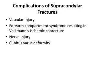
SupracondylarHumerus Fractures:Complications • Compartment syndrome • Vascular injury / compromise • Loss of reduction / Malunion –cubitus varus • Loss of motion • Pin track infection • Neurovascular injury with pin placement
Supracondylar Humerus Fractures- Flexion type • Rare, only 2% • Distal fracture fragment anterior,flexed • Ulnar nerve injury -higher incidence • Reduce with extension • Often requires 2 sets of hands in Or, hold elbow at 90 degrees after reduction to facilitate pinning