Download
1 / 28
280 likes | 383 Views
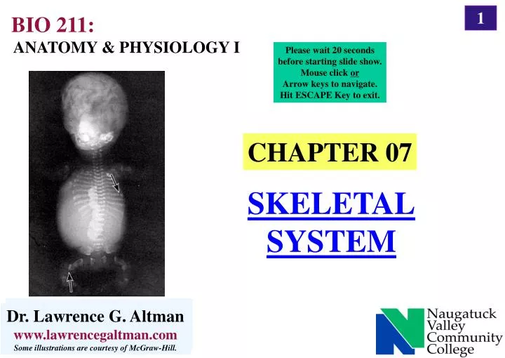
CHAPTER 07. SKELETAL SYSTEM. BIO 211:. ANATOMY & PHYSIOLOGY I. Please wait 20 seconds before starting slide show. Mouse click or Arrow keys to navigate. Hit ESCAPE Key to exit. Dr. Lawrence G. Altman www.lawrencegaltman.com Some illustrations are courtesy of McGraw-Hill.

E N D
CHAPTER 07 SKELETAL SYSTEM BIO 211: ANATOMY & PHYSIOLOGY I Please wait 20 seconds before starting slide show. Mouse click or Arrow keys to navigate. Hit ESCAPE Key to exit. Dr. Lawrence G. Altman www.lawrencegaltman.com Some illustrations are courtesy of McGraw-Hill. Dr. Lawrence G. Altman www.lawrencegaltman.com Some illustrations are courtesy of McGraw-Hill. Dr. Lawrence G. Altman www.lawrencegaltman.com Some illustrations are courtesy of McGraw-Hill. Dr. Lawrence G. Altman www.lawrencegaltman.com Some illustrations are courtesy of McGraw-Hill. Dr. Lawrence G. Altman www.lawrencegaltman.com Some illustrations are courtesy of McGraw-Hill. Dr. Lawrence G. Altman www.lawrencegaltman.com Some illustrations are courtesy of McGraw-Hill. Dr. Lawrence G. Altman www.lawrencegaltman.com Some illustrations are courtesy of McGraw-Hill. Dr. Lawrence G. Altman www.lawrencegaltman.com Some illustrations are courtesy of McGraw-Hill. Dr. Lawrence G. Altman www.lawrencegaltman.com Some illustrations are courtesy of McGraw-Hill. Dr. Lawrence G. Altman www.lawrencegaltman.com Some illustrations are courtesy of McGraw-Hill. Dr. Lawrence G. Altman www.lawrencegaltman.com Some illustrations are courtesy of McGraw-Hill. Dr. Lawrence G. Altman www.lawrencegaltman.com Some illustrations are courtesy of McGraw-Hill.
Bone Function • Body Movement • interacts with muscles • bones act as rigid bar of a lever • Support and Protection • gives shape to head, etc. • supports body’s weight • protects lungs, brain etc. • Inorganic Salt Storage • calcium • phosphate • magnesium • sodium • potassium • Blood Cell Formation • hematopoiesis • red marrow
Bones of the Skeletal System 1. There are 206 bones in the adult; 270 in a newborn. Many fuse during growth and development. 2. The skeleton is divided into axial and appendicular portions. Axial skeleton:the skull, middle-ear bones, the hyoid bone, rib cage, vertebral column, and sternum. Appendicular skeleton:the upper and lower extremities, and the pectoral and pelvic girdles.
Shapes of Bones Long bonesExample: Femur (a) include those in the appendages that produce body movement. Short bonesExample: Tarsal (b) are equal in length and width, such as those of the wrist and ankle. Flat bonesExample: Parietal (c) such as in the skull, protect soft tissues. IrregularExample: Vertebra (d) include the vertebrae and others. SesamoidExample: Patella (e) Round bone with tendons adjacent to joints
General Features of Bones The features of a long bone include its outer layer of compact bone, bone marrow, and spongy bone at its ends. The shaft of a long bone is referred to as the diaphysis;the expanded ends are the epiphyses. 3. The epiphyses are covered with articular cartilage, and the outer bone is covered by periosteum. The inside is lined with endosteum. 4. During growth, an epiphyseal plate of hyaline cartilageforms a model for bone to replace.
Parts of a Long Bone • epiphysis • distal • proximal • diaphysis compactbone spongybone articularcartilage periosteum endosteum • medullary cavity • trabeculae slender plates of spongy bone • marrow- in the spongy bone: • red • yellow
Cells Histology of Osseous Tissue OSTEO = bone Osteogenic cells develop from mesenchyme and occur in the endosteum, the inner periosteum, and in the Haversian canals. They are the only source of new cells of osteoblasts and osteocytes. Osteoblastsare bone-forming cells, and build new bone matrix. Osteocytes are osteoblasts trapped in bone matrix. They remain active in maintenance of bone. Osteoclastsare bone-dissolving cells that form by fusion of monocytes. They break down bone and release its minerals to the blood.
Compact Bone Histology of Osseous Tissue Organicmatter in bone (one-third of the dry weight) collagen, GAGs, proteoglycans and glycoproteins. Mineralcomponentsin bone: especiallyhydroxyapatite and calcium carbonate. Other minerals are present in minute quantities. Lamellae are arranged mostly in concentric circlesaround Haversian canals. This is the basic structural unit of compact bone: osteon. Within the lamellae lie the lacunae with osteocytes. Canaliculi extend between adjacent lamellae. Perforating (Volkmann's) canals enter the bone from the outside and inside, and feed into the Haversian systems, carrying nerves and blood vessels.
Compact Bone (Basic unit) (Haversian) (Haversian) (Volkmann's) Histology of Osseous Tissue
Compact Bone Histology of Osseous Tissue
Marrow Histology of Osseous Tissue • In children, red marrow (myeloid tissue) is hemopoietic and fills the medullary cavity.Myeloid = pertaining to the marrow. Hemopoietic: • adj : pertaining to the formation of blood or blood cells; • "hematopoietic stem cells in bone marrow" [syn: hematopoietic, haematopoietic, haemopoietic, hematotic, haematotic, hematogenetic, haematogenetic] 2. In adults (age 30), most of the marrow in the medullary cavity is yellow marrow that stores fat. 3. In older adults (age 70), most of the yellow marrow isreplaced by gelatinous marrow.
Bone Formation Intramembranous 1. Intramembranous ossification occurs within a membrane of soft tissue that represents the location of a future flat bone. Its cells differentiate into osteogenic cells and osteoblasts, andtrabeculaeare formed. 2. Osteoblasts form on the trabeculae and lay down an organic matrix and deposit calcium phosphate within it. When trapped, they become osteocytes.
Bone Formation Endochondral 1. Endochondral ossification is bone formation using a cartilage model. In the center of the model is the primary ossification center where lacunae enlarge and minerals are deposited around them. 2. The Primary Ossification Center a. Cells of the perichondrium become osteogenic cells and osteoblasts and produce bone on the outside of the model. b. In the center of the model, a primary marrow space is formed. 3. The Metaphysis a. The transition between the head of hyaline cartilage and the primary marrow space is the metaphysis. b. It exhibits five zones representing stages of ossification: the zone of reserve cartilage; the zone of cell proliferation; the zone of cell hypertrophy; the zone of calcification; and the zone of bone deposition.
Bone Formation Endochondral 4. The Secondary Ossification Center a. At birth, secondary ossification centers form in the epiphyses of long bones. The epiphysis is hollowed out from the center outward and is replaced by bone. b. Cartilage remains until adulthood at the epiphyseal plates.
Bone Formation Endochondral
Bone Formation Bone Formation Growth & Remodeling 1. Each year, bone exchanges 18% of its calcium. About 5% of the adult skeleton undergoes remodeling at any one time. Physical activity enlarges bony prominences. 3. Cartilage can grow two ways: by interstitial growth and by appositional growth. 4. In achondroplasticdwarfism, chondrocytes fail to multiply in long bones. Intramembranous vs. Endochondral
Factors Affecting Bone Development, Growth, and Repair Deficiency of Vitamin A retards bone development Deficiency of Vitamin C results in fragile bones Deficiency of Vitamin D rickets: Metabolic Disorder:phosphorous/calciumosteomalacia: bone softening Insufficient Growth Hormone dwarfism Excessive Growth Hormone gigantism Insufficient Thyroid Hormone delays bone growth Sex Hormones promote bone formation; stimulate ossification of epiphyseal plates Physical Stress stimulates bone growth
Physiology Ossesous Tissue Mineral Resorption Resorption is the process of dissolving bone to release its minerals to the bloodstream. Osteoclasts dissolve bone using acid phosphatase. Calcium and Phosphorus Homeostasis The skeleton serves as a reservoir for calcium, phosphorus, and other minerals that play important roles in physiology. Excessively low calcium concentration is called hypocalcemia, causing the nervous system to become hyperexcitable. Muscle tetany can result. Excessive calcium is hypercalcemia, which can cause nervous system depression and sometimes cardiac arrest.
Physiology Osseous Tissue Calcium and Phosphorus Homeostasiscont. The balance between calcium storage (into bone) and calciumresorption (into the blood) is controlled by two hormones: calcitonin acts to lowers blood levels of calcium by stimulating osteoblasts and inhibiting osteoclasts. parathyroid hormone (PTH)raises blood calcium when it drops too low. PTH stimulates osteoclasts, lessens urinary excretion of calcium, and stimulates the synthesis of vitamin D.
Physiology Osseous Tissue Calcium and Phosphorus Homeostasiscont.
Physiology Osseous Tissue Vitamin D Vitamin D is a hormone that is produced in concert by the skin, liver, and kidney. The most active form is calcitrol, produced together by the skin (with UV light), liver, and kidney. Calcitrol promotes intestinal absorption of calcium and phosphate while reducing urinary elimination of these minerals. Insufficient vitamin D can causerickets in children and osteomalacia in adults. (see previous chart)
Bone Disorders Types of Fractures
Bone Disorders Fractures and their Repair The Healing of Fractures A bone fracture results in a hematoma from torn blood vessels. Next, soft granulation tissue forms as blood vessels grow into the hematoma. Macrophages remove debris as osteoclasts, osteogenic cells; fibroblasts migrate to the area. Fibroblasts deposit collagen, and a fibrocartilage callus is formed by chondroblasts. The callus is first soft, then hard as it is replaced with bony tissue. The area of the fractureis remodeled for 3-4 months until broken bone fragments are resorbed.
Bone Disorders Fractures and their Repair
Bone Disorders Treatment of Fractures Fractures may be set by: closed reduction no surgery open reduction surgical placement of bones, using pins and plates. Orthopedics: branch of medicine dealing with injuries/disorders of bones, joints, and muscles.
Bone Disorders Osteoporosis The most common bone disease is osteoporosis in which bones lose mass and become brittle. The group most prone to this disease are elderly, postmenopausal white women; black women are rarely afflicted. The spine commonly becomes compressed, a condition leading to kyphosis. Disuse osteoporosis occurs at any age due to immobilization or inadequate weight-bearing exercise.
LAST SLIDE