Download
1 / 2
20 likes | 82 Views
This study presents findings on gamma response to various stimuli in depth-electrode recordings. It includes data on trial-to-trial gamma power responses and detailed analysis of gamma amplitude to different stimuli. The localization of the recording electrode and specific responses to letter-strings are highlighted, offering insights into brain activity patterns. The study sheds light on neural responses to target images, exclusion criteria, and electrode contact analyses.

E N D
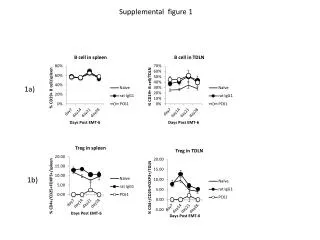
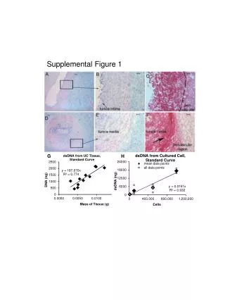
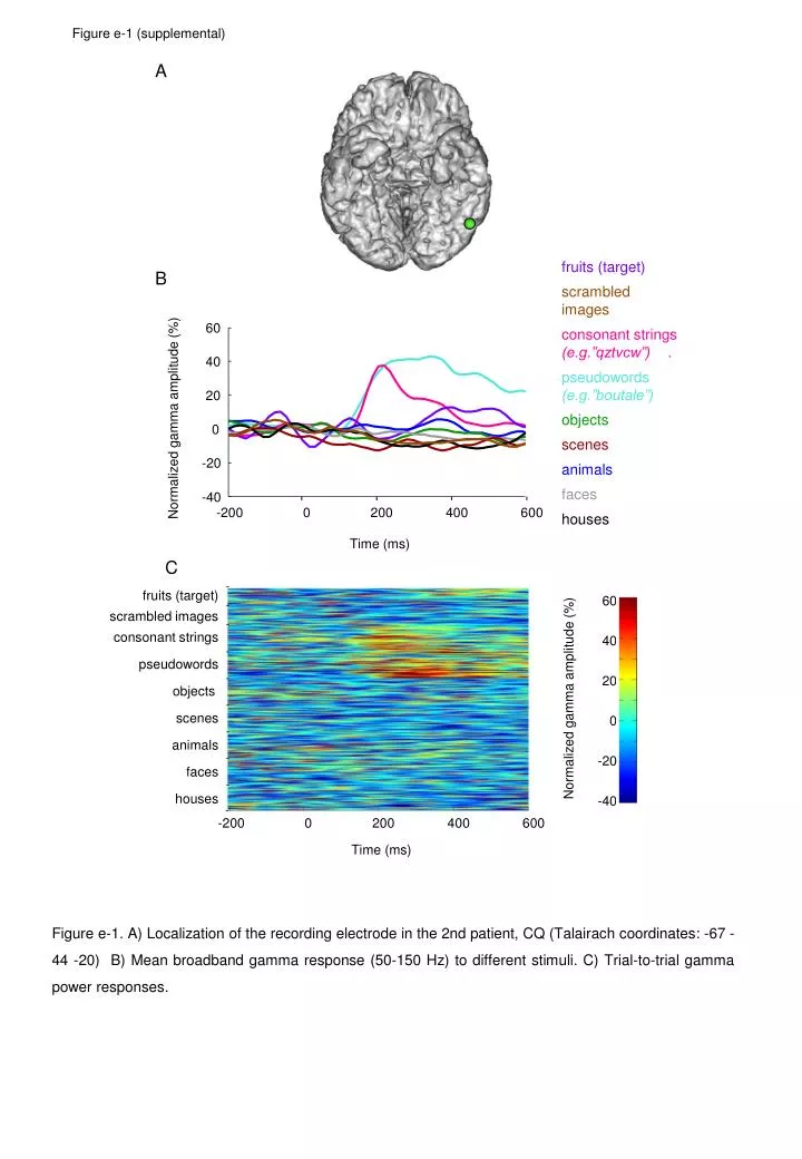
-200 0 200 400 600 Time (ms) Figure e-1 (supplemental) A fruits (target) B scrambled images 60 consonant strings (e.g.”qztvcw”) . 40 pseudowords (e.g.”boutale”) 20 Normalized gamma amplitude (%) objects 0 scenes -20 animals faces -40 -200 0 200 400 600 houses Time (ms) C fruits (target) 60 scrambled images consonant strings 40 pseudowords 20 objects Normalized gamma amplitude (%) scenes 0 animals -20 faces houses -40 Figure e-1. A) Localization of the recording electrode in the 2nd patient,CQ (Talairach coordinates: -67 -44 -20) B) Mean broadband gamma response (50-150 Hz) to different stimuli. C) Trial-to-trial gamma power responses.
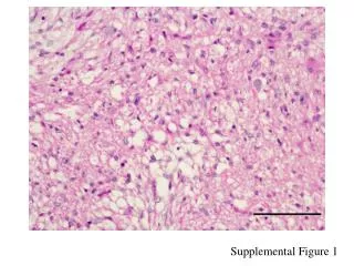
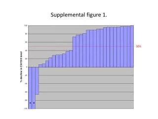
Figure e-2 (supplemental) A z=-10 y=-57 y=-57 B gamma amplitude (0-200 ms) to pseudowords normalized to largest non-character response (%) 500 250 0 pseudo- words pseudo- words pseudo- words strongest response to: animals animals electrode contacts 2 3 7 8 9 3.5 mm Figure e-2. Specific responses to letter-strings are confined to one electrode contact only. A) Structural T1-weigthed MRI image of subject PM with implanted depth-electrodes. The depth-electrode of interest is highlited by a white rectangle. B) Gamma amplitude responses to pseudowords at neighbouring electrode contacts along the depth-electrode. Responses were normailzed to the largest non-character response; for example, at electrode contact 7, 100% is equal response to animals, the strongest non-character response at that electrode. Note that specific responses to letter-strings drop off sharply from over 500% at contact 7 to 109% at contact 8. Responses of contact 7 are depicted in detail in Figure 1. Gamma response amplitudes were calculated in a 0-200 ms window. Responses to target images (fruits) were excluded from the figure to avoid contamination with top-down effects and motor preparation. Electrode contacts with poor signal or no signal are omitted. Note that the size of the electodes in A) (0.8 mm diameter) is magnified by the MRI artefact.