Download
1 / 23
240 likes | 411 Views
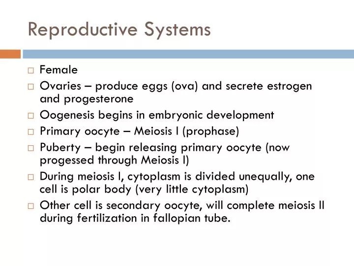
Reproductive Systems. Female Ovaries – produce eggs (ova) and secrete estrogen and progesterone Oogenesis begins in embryonic development Primary oocyte – Meiosis I (prophase) Puberty – begin releasing primary oocyte (now progessed through Meiosis I)

E N D
Reproductive Systems • Female • Ovaries – produce eggs (ova) and secrete estrogen and progesterone • Oogenesis begins in embryonic development • Primary oocyte – Meiosis I (prophase) • Puberty – begin releasing primary oocyte (now progessed through Meiosis I) • During meiosis I, cytoplasm is divided unequally, one cell is polar body (very little cytoplasm) • Other cell is secondary oocyte, will complete meiosis II during fertilization in fallopian tube.
Suspensory ligament of ovary Uterine (fallopian) tube Ovarian blood vessels Fundus of uterus Lumen (cavity) of uterus Ovary Infundibulum Broad ligament Uterine tube Fimbriae Ovarian ligament Round ligament of uterus Body of uterus Endometrium Wall of uterus Myometrium Perimetrium Ureter Cervical canal Uterine blood vessels Uterosacral ligament Vagina Cervix (b) Figure 16.8b
Menstrual CyclePhase 1 Follicular phase (10 days) • Anterior pituitary secretes FSH and LH • FSH – follicle stimulating hormone • LH – luteinizing hormone • Follicle grows – releases estrogen which causes uterine lining to thicken-endometrium • Estrogen also causes ant. Pit. To release LH • The luteal surge causes ovulation
Growing follicles Primary follicle Degenerating corpus luteum Blood vessels Antrum Corona radiata Mature vesicular (Graafian) follicle Germinal epithelium Corpus luteum Developing corpus luteum Ruptured follicle Ovulation Secondary oocyte Figure 16.7
Ovulation – release of the egg from the ovary ends the follicular phase. • Ova floats into fallopian tube (oviduct)
Phase 2 Luteal phase • Follicle has released the egg and changes into the corpus luteum • Corpus luteum continues to secrete estrogen and now begins to produce progesterone • Progesterone increases vessels and glands in uterus • After about 13-15 days, if fertilization and implantation have NOT occurred the corpus luteum shuts down
Phase 3 Menstruation • Uterus reabsorbs some of the tissue created in the endometrium • The rest sheds off and is passed out of the body
Pregnancy • If pregnancy occurs, the fetus releases HGC human chorionic gonadotropin • This maintains the uterine lining
Male Reproductive system Ureter Urinary bladder Seminal vesicle Prostatic urethra Ampulla of ductus deferens Pubis Membranous urethra Ejaculatory duct Urogenital diaphragm Rectum Erectile tissue of the penis Prostate Bulbourethral gland Spongy urethra Shaft of the penis Ductus (vas) deferens Glans penis Epididymis Testis Prepuce (a) Scrotum External urethral orifice Figure 16.2a
Male reproductive organs • Sperm undergo meiosis I and II in the seminiferous tubules (primary to secondary spermatocyte) • Mature in the epididymis (secondary spermatocyte becomes spermatazoa) • Seminal vesicles produce fructose fluid for energy • Prostate secretes alkaline substance to neutralize vaginal fluids • FSH – stimulates sperm production • LH – stimulates interstitial cells to produce testosterone
Development • Morphogenesis – organization of human structure • When an egg is fertilized it becomes the zygote • Cleavage – rapid cell division • Solid ball of cells – morula • Next stage is the blastula – cells divide and press close to each other leaving a fluid-filled cavity called a blastocoel • Cells migrate into the blastocoel forming the gastrula • Endoderm, mesoderm, ectoderm form
Indeterminate and Determinate Cleavage • Indeterminate – if daughter cells are separated, they can individually complete normal development. • Determinate cells cannot
Egg Cytoplasm • Cytoplasmic material is unequally distributed in the egg, cytoplasmic material will vary in daughter cells • This results in embryonic axes • Gray crescent in frog eggs – region essential in frog development • Morphogens – proteins with asymmetrical distribution in oocyte – determines dorsal/ventral side, anterior/posterior regions of the cell, also act as transcription factors
Embryonic Induction • Organizers are cells that exert influence over other cells during development through cell-cell communication. Regulates the expression of certain genes • Dorsal lip of the blastopore in a newt is an organizer – produces a notochord • In a roundworm, the anchor cell secretes an inducer molecule that binds to pm of neighboring cells, activate trans. Factors, they bind to promoter on DNA, signal the synthesis of a protein that stimulates the differentiation into a vulva cell.
Apoptosis • Programmed cell death • External signal initiates a signal trans. Pathway, activating cell death genes (Ced genes in C. elegans), destroys 131 different cells. • Humans have webbing during development. Activation of apoptosis of certain cells, cause fingers and toes to separate.
Control of Gene expression • Methylation of histones in chromatin, prevents expression • microRNAs – small segments of RNA that influence expression by blocking mRNA translation • Homeotic genes – master genes that specify development. Contain homeobox, Hox genes • Hox genes code for transcription factors, dictate the spatial organization of fruit flys
Extra-embryonic membranes • Yolk sac, amnion, allantois, chorion