Download
1 / 15
160 likes | 207 Views
Explore the acoustic reflex, a protective function in response to intense sound, and learn about the inner ear anatomy including the cochlea and vestibular system.

E N D
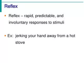
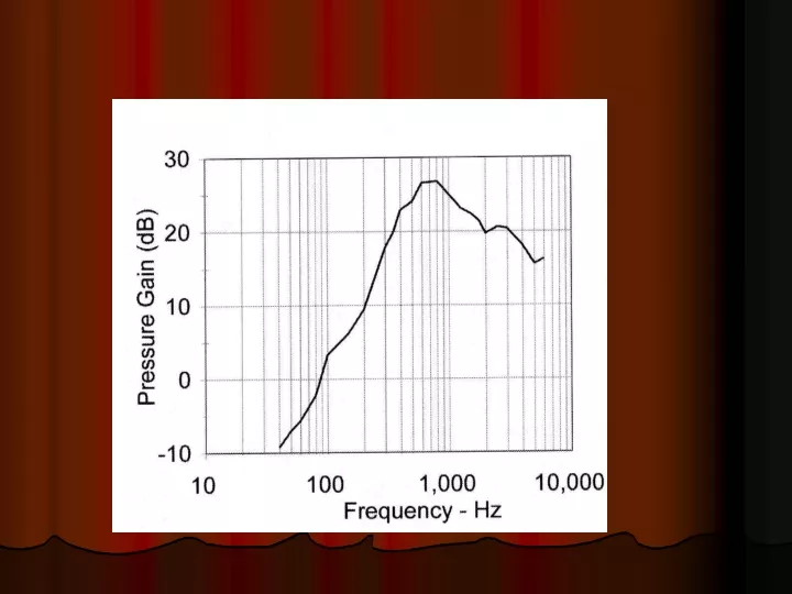
Acoustic reflex Protective function Due to muscle contraction in response to intense sound Threshold of reflex: Around 80 dB SL (sensation level). Reflex results in attenuation of loud sounds by about 10-30 dB More effective at low frequencies (less than 2 kHz) Not very effective for short duration sounds: Latency about 10-150ms
Measurement of acoustic reflex Middle ear muscles contract: Stiffness increases More sound reflected back Lack of acoustic reflex: Implies problem in middle ear muscles/part(s) of acoustic reflex pathway
Inner ear Series of interconnecting canals or ‘labyrinths’ in the temporal bone Two types: Osseous Bony Bigger cross-sectional area Contains fluid called perilymph Membraneous Soft tissue Situated within the bony labyrinth Contains fluid called endolymph http://research.meei.harvard.edu/Otopathology/3dmodels/download.html
Labyrinths contain Three parts Semicircular canals Vestibular system Vestibule Cochlea Auditory system
Vestibular system Function: Balance and equilibrium Sensory cells: Hair cells
Auditory system: Cochlea Name derived from snail-like shape Tube of decreasing diameter coiled around itself Coiled around a central bony canal called modiolus Broad base, narrow apex Length: About 35 mm In human beings: 2 and 5/8 coils
Isolated cochlear turns from the inner ear of fetal sheep: Photo courtesy of Gerhardt, K.
Cochlear macrostructure Partially divided throughout its length by a thin spiral shelf of bone called osseous spiral lamina At the outer (lateral) wall of the cochlea: Spiral ligament
Between the osseous spiral lamina and the spiral ligament: Basilar membrane Runs all the way along the length of the cochlea, except for a small opening at the apex called the helicotrema Reissner’s membrane projects diagonally from spiral lamina to outer bony wall of cochlea Joins the basilar membrane at the helicotrema
Between the osseous spiral lamina and the spiral ligament: Basilar membrane Runs all the way along the length of the cochlea, except for a small opening at the apex called the helicotrema Reissner’s membrane projects diagonally from spiral lamina to outer bony wall of cochlea Joins the basilar membrane at the helicotrema http://www.iurc.montp.inserm.fr/cric/audition/english/ear/fear.htm http://epl.meei.harvard.edu/~hwang/3Dviewer/3Dviewer.html
Cross-section of the cochlea Basilar membrane and Reissner’s membrane divide the cochlear canal into three ducts: Scala vestibuli Scala tympani Scala media
Scala Vestibuli Above Reissner’s membrane Extends from oval window in the vestibule to the helicotrema Contains perilymph
Scala Tympani Below the basilar membrane Extends from round window to the helicotrema Contains perilymph
Scala media/cochlear sac Bound below by basilar membrane Bound above by Reissner’s membrane Bound on the outer side by Stria vascularis Contains endolymph