Download
1 / 69
700 likes | 1k Views
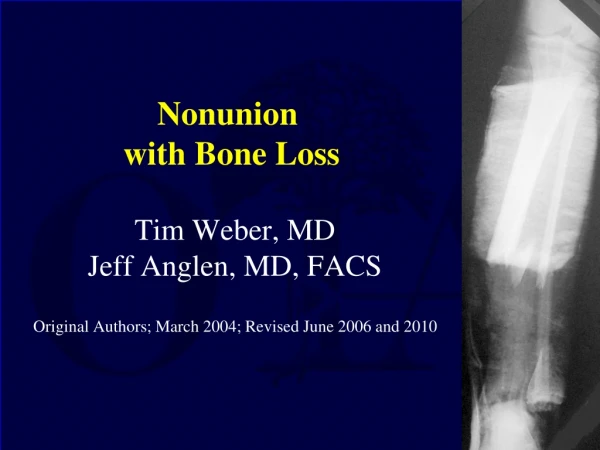
Nonunion with Bone Loss. Jeff Anglen, MD, FACS Professor and Chairman, Department of Orthopaedics Indiana University Created March 2004; Revised June 2006. Etiology. Open fracture segmental post debridement blast injury Infection Tumor resection Osteonecrosis. Classification.

E N D
Nonunion with Bone Loss Jeff Anglen, MD, FACS Professor and Chairman, Department of Orthopaedics Indiana University Created March 2004; Revised June 2006
Etiology • Open fracture • segmental • post debridement • blast injury • Infection • Tumor resection • Osteonecrosis
Classification Salai et al. Arch Orthop Trauma Surg 119
Classification Not Widely Used Not Validated Not Predictive Salai et al. Arch Orthop Trauma Surg 119
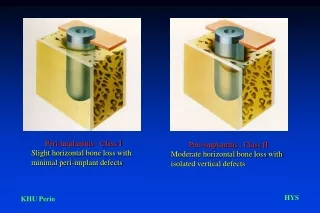
Evaluation • Soft tissue envelope • Infection • Joint contracture and range of motion • Nerve function: sensation, motor • Vasculature: perfusion, angiogram? • Location and size of defect • Hardware • General health of the host • Psychosocial resources
Is it Salvageable? • Vascularity - warm ischemia time • Intact sensation • other injuries • Host health • magnitude of reconstructive effort vs patient’s tolerance • ultimate functional outcome
Priorities • Resuscitate • Restore blood supply • Remove dead or infected tissue (Adequate debridement) • Restore soft tissue envelope integrity • Restore skeletal stability • Rehabilitation
Bone Loss - Initial Treatment • Irrigation and Debridement
Bone Loss - Initial Treatment • Irrigation and Debridement • External fixation
Bone Loss - Initial Treatment • Irrigation and Debridement • External fixation • Antibiotic bead spacers
Bone Loss - Initial Treatment • Irrigation and Debridement • External fixation • Antibiotic bead spacers • Soft tissue coverage
Bone Loss - Initial Treatment • Irrigation and Debridement • External fixation • Antibiotic bead spacers • Soft tissue coverage • Sterilization and Re-implantation?
Potential Segment Re-implantation • Young, healthy patient • well vascularized soft tissue bed (femur, not tibia) • single cleanable fragment • early, aggressive, meticulous wound care • adequate sterilization of the fragment • Antibiotics, local and systemic Mazurek et al J. Ortho Trauma 2003
Skeletal Stability: Treatment Options • Significant loss of joint surface • osteochondral allograft • total joint or hemi- arthroplasty • arthrodesis
Autogenous bone graft cancellous cortical vascularized Allogeneic bone graft cancellous cortical DBM Distraction osteogenesis multifocal shortening/ lengthening bone transport Salvage procedures shortening one bone forearm Skeletal Stability: Treatment Options for Diaphyseal Defects
Bone Grafting • Osteogenesis - bone formation 1. Survival and proliferation of graft cells 2. Osteoinduction - host mesenchymal cells • Osteoconduction • Structural Support
Graft Incorporation • Hemorrhage • Inflammation • Vascular invasion • Osteoclastic resorbtion/ Osteoblastic apposition • Remodelling and reorientation
Autogenous Cancellous Bone Grafting • Quickest, highest success rate • little structural support • best in well vascularized bed • donor site morbidity • quantity limited - short defects?
Papineau Technique • Direct open cancellous grafting of granulation bed • typically large metaphyseal defect
22 yo man • RHD • MCA • open segmental humerus fracture with bone loss and radial nerve out
Irrigation and Debridement Application of external fixator Wound care Antibiotics
Posterior plate fixation Iliac crest bone grafting + antibiotic CaSo4 beads Implantable bone stimulator
40 yo female 10 years after cancellous grafting of distal tibial defect
Allograft • Incorporates like autograft, but slower • No cells survive • may include joint • No size or quantity limitation • risk of disease transmission • infection rate ~ 5-12% • Intercalary grafts for tumor resection >80% success (Ortiz-Cruz, et al.) • can be combined with autograft
Cortical Strut Grafting • Provide structural support • weakly osteogenic • revascularize slowly • initially become weaker • frequently needs supplementary cancellous graft for union(Enneking, JBJS 62-A, 1980)
35 yo MVC Open femur with segmental bone loss I&D ExFix Beads
ORIF with bladeplate fibular strut allograft cancellous autograft CaSO4 pellets Bone stimulator
8 months FWB without pain return to work
Cancellous Allograft • May be similar to cancellous AUTOgraft when combined with recombinant human bone morphogenic protein (rhBMP) or other growth factors • Cook et al. Evaluation of INFUSE Bone Graft in a Canine Critical Size Defect: Effect of Sponge Placement on Healing, OTA annual meeting 2005 http://www.hwbf.org/ota/am/ota05/otapa/OTA050936.htm • Volgas and Stannard, A Randomized Controlled Prospective Trial of Autologous Bone Graft versus Iliac Crest Bone Graft for Nonunions and Delayed Unions , OTA annual meeting 2004 http://www.hwbf.org/ota/am/ota04/otapa/OTA041165.htm
Vascularized Graft • Pedicled ipsilateral fibula • Free bone flap • fibula • iliac crest • rib • Structural support, rapid healing, independent of host bed • will hypertrophy
The Free Fibula • Taylor 1975 • branch of the peroneal and periosteal vessels • Can be transferred with skin or with skin and muscle to reconstruct several tissues at once (Jupiter et al., Heitmann et al.) • donor site morbidity • mod. Gait changes up to 18 months • sl. calf strength, eversion • FHL contracture • peroneal paresthesias
29 yo RHD female GSW L arm Pulses intact Hand neuro exam intact
Irrigation Debridement ExFix wound care
5 months Free fibula graft fixation with long T plate
21 mon. 10 mon. 14 mon.
24 months post injury revision fixation proximally with bone graft
3 years post- injury healed uses hand for ADLs
40 yo female 10 years after free fibula graft for femoral defect Hypertrophy and consolidation
Distraction Osteogenesis • Ilizarov 1951 “tension-stress effect” • mechanical induction of new bone formation • neovascularization • stimulation of biosynthetic activity • activation and recruitment of osteoprogenitor cells • intramembranous ossification
Ilizarov Technique • Rings and Tensioned wires • corticotomy • latency period • gradual distraction, .25 mm q60 • parallel fibrovascular interface • columns of ossification
Ilizarov Technique • Acute shortening and compression at fracture site, followed by lengthening at a separate site • reduces soft tissue defect • protects vascular/nerve repair • Bone Transport - internal lengthening of one or both segments to fill gap • allows normal length and alignment during treatment
High rate of ultimate success, good restoration of length and alignment No donor site morbidity May be functional during treatment Requires prolonged time in the frame ~ 2 mon/cm frequent docking site problems requiring bone grafting frequent complications Bone Transport But... Transport over an IM nail (Monorail technique) or under a MIPO plate
25 yo ♀ AK-47 GSW This case and images courtesy of Kevin Pugh, MD Ohio State University
Irrigation Debridement External Fixation This case and images courtesy of Kevin Pugh, MD Ohio State University
Application of circular frame with half-pins for transport This case and images courtesy of Kevin Pugh, MD Ohio State University
Retrograde transport of a 14 cm segment required 2 years in the frame This case and images courtesy of Kevin Pugh, MD Ohio State University