Download
1 / 26
310 likes | 772 Views
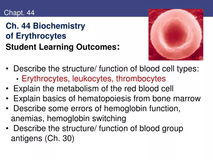
Chapt. 44. Ch. 44 Biochemistry of Erythrocytes Student Learning Outcomes : Describe the structure/ function of blood cell types: Erythrocytes, leukocytes, thrombocytes Explain the metabolism of the red blood cell Explain basics of hematopoiesis from bone marrow

E N D
Chapt. 44 • Ch. 44 Biochemistry • of Erythrocytes • Student Learning Outcomes: • Describe the structure/ function of blood cell types: • Erythrocytes, leukocytes, thrombocytes • Explain the metabolism of the red blood cell • Explain basics of hematopoiesis from bone marrow • Describe some errors of hemoglobin function, anemias, hemoglobin switching • Describe the structure/ function of blood group antigens (Ch. 30)
Blood cells • Table 1 Blood cells (cells/mm3): • Erythrocytes 5.2 x 10⁶ men • carry oxygen 4.6 x 10⁶ women • Neutrophils 4300 • granules; phagocytic, O2 burst kills • Lymphocytes 2700 • immune response, B- and T-cells, NK • Monocytes 500 • macrophages for bacteria, damage • Eosinophils 230 • granules destroy parasites (worms) • Basophils 40 • granules hypersensitivity, allergic histamine, proteases,
Hematopoiesis • Hematopoiesis: • Stem cells in bone marrow (1/105) • Proliferate, differentiate, mature • by growth factors, hormones • signal transduction paths • Myeloid, lymphoid lines • Leukemias: immature cells • keep proliferating; • defined by cell type Fig. 15
Anemia • Anemias: hemoglobin concentration is low: • Normal Hb g/dL: men 13.5-17.5; women 11.5-15.5 • Anemias classified by red blood cell morphology: • Rbc morphology functional deficit possible cause • Microcytic, impaired Hb thalassemia, lead, • hypochromic synthesis iron deficiency • Macrocytic impaired DNA vit B12 or folic acid • normochromic synthesis deficient, erythroleukemia • Normocytic red cell loss acute bleeding, • normochromic sickle cell defects
Erythrocyte metabolism • Erythrocyte metabolism: Only glycolysis • ATP for Na+/K+, Ca2+ • HMP shunt makes NADPH • G6PD is 1st enzyme • Lifetime rbc by G6PD activity • 2,3-BPG modulates O2 binding • Need Fe2+ Hb bind O2; • If ROS made Fe3+, NADH can reduce Fig. 1
Heme synthesis • Heme synthesis in erythrocyte precursor: • Heme = porphryn ring, coordinated to Fe • Complexed to proteins in hemoglobin, myoglobin and cytochromes; most common porphryn in body • 4 pyrrole rings with –CH- joining • Various side chains • Heme is red color Fig. 2
Heme synthesis • Heme synthesis: • Glycine, succinyl CoAform • d-Aminolevulinic acid • (d-ALA) • Each heme needs 8 of each • Final step is Fe2+ • Heme regulates: • inhibit 1st enzyme • repress synthesis • Porphyria diseases from • defective enzymes • intermediates accumulate • photosensitive, toxic products Fig. 3
Heme synthesis • Heme synthesis begins with d-ALA: • Decarboxylation by d-ALA synthase • PLP is pyridoxal phosphate • Dehydratase joins 2 d-ALA • 4 pyrroles form porphyrinogen Fig. 4
Sources of iron and heme • Iron is essential from diet – 10-15 mg/day recommended • Iron is not readily absorbed from many sources • Iron in meats is form of heme, readily absorbed • Nonheme iron of plants not as easily absorbed becauuse other compounds precipitate iron • Iron absorbed in ferrous state (Fe2+), oxidized by ferroxidase to Fe3+ for transport • Apotransferrin binds Fe3+ = Transferrin • Stored as ferritin in cells • Heme stimulates synthesis of globin proteins from ribosomes
Iron metabolism • Iron metabolism: • Transferrin carries Fe3+ to cells; stored as ferritin • Transferrin taken up by R-mediated endocytosis • Hemosiderin stores excess Fig. 6 RE = reticulo-endothelial system
Degradation of hemoglobin • Heme is degraded to bilirubin: • Bilirubin is congugated to glucuronate (more soluble),excreted • Rbc only live ~120 days • Globin is degraded to amino acids Figs. 7,8
Red blood cells • Erythrocyte cell membrane: • Red disc, pale center • Biconcave shape • Maximizes surface area • 140 um2 vs. 98 um2 sphere • Deforms to enter tissues • Spleen destroys damaged • red blood cells Fig. 9
Cytoskeleton of erythrocyte • Erythrocyte cytoskeleton • provides shape, structure, permits stretch • 2-D lattice of proteins links to membrane proteins: • spectrin (a, b) • actin • ankyrin • band 4.1 • membrane proteins: • glycophorin • band 3 protein • Mature rbc does not synthesize • new proteins • Gets lipids from circulating LDL Fig. 10 general side view; inside cell view up
Agents affect oxygen binding of hemoglobin • Agents affect oxygen binding of hemoglobin: • 2, 3-BPG (glycolysis intermediate) binds between 4 subunits of Hb, lowers affinity for O2, releases O2 to tissues • Proton (Bohr) effect: ↑H+ lowers affinity of Hb for O2: • CO2 can bind to Hb (not only bicarbonate) Fig. 11,12, 14
Effect of H+ on oxygen binding to Hb • Effect of H+ on oxygen binding to Hemoglobin: • Tissues: CO2 released → carbonic acid, H+ • H+ bind Hb → release O2 to tissues • Lungs reverse: O2 binds H+Hb → release H+ • H2CO3 forms, releases CO2 to exhale Fig. 13
Hematopoiesis • Hematopoiesis: • Stem cells in bone marrow • proliferate • differentiate • mature • myeloid vs. lymphoid • Stromal cells secrete • growth factors • Cytokines signal via • membrane receptors Fig. 15
Bone marrow • Bone marrow stromal cells secrete growth factors • Hematopoietc stem cells respond
Hematopoiesis involves cytokine signaling • Growth factors signal through membrane receptors: • Ligand causes receptors to aggregate • Activates JAK (kinases) by phosphorylation (cytoplasmic RTK) • JAK phophorylates cytokine receptor on Tyr • Other signaling molecules bind, including STAT (signal transducer and activator of transcription) → nucleus transcription • Also RAS/Raf/MAP kinase activated • Overactive signal → cancer • Transient signal: SOCS silences Figs. 16; 11.15
Erythropoiesis • Erythropoiesis: • Erythropoietin from kidney increases red blood cell proliferation (if low oxygen) • Reticulocytes still have ribosomes, mRNA to make Hb • Mature in spleen, lose ribosomes • Make 1012 rbc/day • Anemia if not • appropriate diet • Iron, vitamin B12, folate Fig. 17
Hemoglobin genes • Hemoglobinopathies, hemoglobin switching: • Order of genes parallels development, controls • >700 mutant Hb (often base subsittution) • HbS sickle cell (Hb b Glu6Val) • HbC (Hb b Glu6Lys) • Both ↑ malaria resistance Fig. 18
Thalassemias • Thalassemias: unequal production of a, b of Hb: • need a:b 1:1 • a has 2 genes each chromosome; b only 1 • can have amino acid substitutions, promoter mutations, gene deletions, splice • Improper synthesis cause instability, or aggregation • b+ has some b; b0 makes none • People offten survive if hereditary persistence of fetal hemoglobin: HPFH (a2g2 = HbF) • Treatments of b-thalassemia or sickle cell: • increase Hb g transcription
VI. Hemoglobin switching • Hemoglobin switching: • embryo blast synthesis yolk • fetus liver synthesis • adult bone marrow • Multiple genes for Hb • Order of genes parallels development • Problems if deletions, other mutations • Problems if imbalance Fig. 18
Transcription factors control Hb switching • a-globin locus about 100 kb; HS40 control region • b-globin locus has LCR control region • Promoter of g gene has many transcription factors that bind; HPFH mutations often map promoter • Mutated repressor (CDP) or site • SSP and SP1 compete for binding near TATA Fig. 19
Blood types reflect erythrocyte glycolipids • Blood group substances are glycolipids or glycoproteins on cell surface of erythrocytes: • Glycosyltransferases add sugars, detemine blood type • Two alleles (three choices) iA, iB, i • Produced in Golgi, lipid part of membrane of vesicle, fuses and carbohydrate extends extracellular Fig. 30.16,17
Key concepts • Key concepts: • Blood contains distinct cell types • Erythrocytes transport O2 and return CO2 to lung • Limited metabolism • Heme synthesis in rbc precursos • Oxygen binding • Hematopoiesis from bone marrow • Leukocytes include monocytes, polymorphonuclear • Hemoglobin mutant proteins, expression
Review question • Review question: • A compensatory mechanism to allow adequate oxygen delivery to tissues at high altitudes, where oxygen concentrations are low, is which of the following? • Increase in 2,3-bisphosphoglycerate synthesis by rbc • Decrease in 2,3-bisphosphoglycerate synthesis by rbc • Increase in hemoglobin synthesis by rbc • Decrease in hemoglobin synthesis by rbc • Decreasing the blood pH