Download
1 / 59
590 likes | 609 Views
Explore the intricate stages of brain and spine formation from embryonic development to myelination and neural tube histology. Covers primary and secondary neurulation, germinal matrix, neuronal migration, and more. Discover the essential steps of neurodevelopment through detailed descriptions and illustrations.

E N D
embryology of BRAIN & SPINE saRavanan
overview Dorsal induction Ventral induction Neuronal prolif.,diff.&histogenesis Neuronal migration Axonal myelination
dorsal induction Formation of neural plate, notochord, neural groove, neural fold and neural tube 4 – 7 wks Primary neurulation Secondary neurulation
ventral induction Formation of brain vesicles, telencephalon, diencephalon, mesencephalon, metencephalon, myelencephalon 5 – 10 wks
neuronal prolif. , diff. & histogenesis Germinal matrix formation, prolif of neurons & differentiation, choroid plexus formation and CSF formation 2 – 5 months and after birth
neuronal migration from ventricular, subventricular layers around primitive brain vesicles to supf cortex,deep nuc of cerebrum & cerebellum 3,4,5 months formation of corpus callosum, commissures, interhemispheric neuronal migrations
axonal myelination Starts at brainstem, cerebellum, thalamus, internal capsule Other areas after birth 3wks – 2yrs and into adolescence
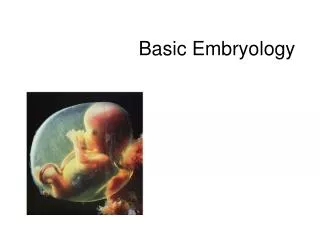
morulation 2 days 3 days 2 cell stage 4 cell stage morula
blastocyst formation 4 days embryoblast blastocele trophoblast
bilaminar disc 8 days Amniogenic cells Amniotic cavity Ectoderm (columnar) Endoderm (cubical) Trophoblastic cells Yolk sac
gastrulation 16 days Prochordal plate (cubical->columnar endoderm)
gastrulation 17 days Primitive streak (prolif of ectoderm)
gastrulation 19 days Mesodermal prolif (intra embryonic)
gastrulation (trilaminar disc) 19 days Prochordal plate (cubical->columnar endoderm) Mesodermal prolif (intra embryonic) Primitive streak (prolif of ectoderm)
Notochord formation 20 days Prochordal plate Notochordal process Primitive knot/primitive node/ HENSON’s node Primitive streak elongates Cloacal memb
neural tube formation 21-23 days Notochord guides NT formation Ectoderm overlying notochord from prochordal plate to primitive node Primary Neurulation- from neural plate, cranial end to L1,L2 Secondary Neurulation- lower L,S,Co from caudal cell mass; no neural plate
neural tube formation Neural groove <Median Hinge Protein> Elevation&Convergence <DorsoLateral Hinge Protein> Closure: middle- cranial(25 day)- caudal(27 day) Neural crest from edge of neural plate to dorsum of Neural tube Dysjn. of cutaneous & neural ectoderm
somites Intraemb mesoderm-paraxial,lateral plate,intermed Paraxial mesoderm segmented to form SOMITES on either side of neural tube --dorsolateral Dermatomyotome: skeletal muscle, dermis --ventromedial Sclerotome: cartilage, bone, ligaments of vertebral column
cranial neuropore somites caudal neuropore
Neural Tube- CNS (brain & spinal cord) Neural Crest- PNS & ANS Somites- skull, vertebra, ligaments, muscle Notochord- induces neurectoderm- Neural tube and deriv. mesenchyme- spinal column, ligaments, muscles remains nucleus pulposus
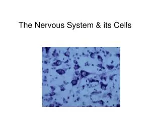
neural tube histology Matrix layer/ependymal/germinal layer- nerve cells, glial cells & more germinal cells produced Mantle layer- developing nerve cells & glial cells Marginal layer- no nerve cells; reticulum of glial cells into which developing nerve cells grow
cells Neuroblasts (apolar-bipolar- multipolar-dendrites formation-synapse formation) neurons Glioblasts/Medulloblasts astroblastsastrocytes oligodendroblastsoligodendrocytes Mesodermal (migrates along with blood vessels) microglia
myelination PNS Schwann cells (from neural crest)invaginates around axon and forms multiple layers into which lipids are deposited CNS Oligodendrocytes form myelin sheath
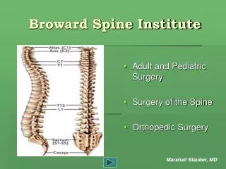
spinal cord dorsal part grows faster,thicker,forms- basal lamina- motor in function alar lamina- sensory in function divided by sulcus limitans dorsal nerve roots are formed from neural crest Recession of spinal cord due to relative differential growth
medulla From Myelencephalon Roof plate widens Sulcuslimitansdivides dorsal(lateral) alar lamina- caudal bulbopontine extension olivary nuclei Sensory nuclei of cranial n of medulla ventral(medial) basal lamina- Motor nuclei of cranial n of medulla
pons From ventral part of Metencephalon Cranial bulbopontine extension pontine nuclei Axons of bulbopontine extension Middle cerebellar peduncle Alar and basal lamina cranial n nuclei Fibres from cortex corticospinal, corticopontine& corticobulbar tracts Lateralpart of alar lamina rhombic lip (cerebellum)
midbrain From mesencephalon Dorsal or alar lamina oculomotor n nuc trochlear n nuc Edinger Westphal nuc Ventral or basal lamina cells of colliculi red nucleus substantia nigra Descending fibres form crus cerebri/basis pedunculi
cerebellum From dorsolateral part of alar lamina of Metencephalon Cells migrate from mantle -marginal layer Cortex Cells that do not migrate dentate, emboliform, globose& fastigial nuclei Supr cerebellar peduncle- axons out of dentate nucleus Middle- axons from pontine nuclei into cerebellum Inferior- axons from spinal cord & medulla
cerebrum fromtwo telencephalic vesicles of prosencephalon Forms together with Corpus striatum Grows upward, forward, backwards.. Encloses lateral ventricles within Medial wall invaginates – choroid fissure Fold of pia extends in-- telachoroidea Tuft of capillaries enter– choroid plexus
thalamus&hypothalamus From diencephalon Two grooves— epithalamic sulcus hypothalamic sulcus Forms three regions— epithalamus(habenular nuc&pineal body) thalamus hypothalamus
neural crest derivatives Neurons of spinal postr nerve root ganglion Neurons of sensory ganglia of V,VII,VIII.IX,X Neurons of Sympathetic ganglia Schwann cells of all peripheral nerves Cells of adrenal medulla Chromaffn tissue Melanoblasts of skin
ANS Sympathetic preganglionic– mantle region of T1 – L2,3 postganglionic– neural crest Parasympathtic preganglionic– cranial- GVE brainstem- EW nuc, salivary, lacrimatory, dorsal nuc of X sacral- mantle layer of sacral spinal cord postganglionic-- same regions
skull -viscerocranium(from neural crest) bones of face -neurocranium(from mesoderm of occipital somites) --membranous part vault of skull with flat bones separated by sutures --cartilaginous part base of skull wth many ossific centres Postr fontanelle closes by 2 months Antr fontanelle closes by 2 years Defects: anencephaly, scaphocephaly, plagiocephaly,microcephaly, mandibulo facial dysostosis, cong hydrocephalus
vertebra From sclerotomes of somites Sclerotome- 3 parts; cranial, middle, caudal Vertebra- fusion of caudal part of upper & cranial part of lower sclerotome, so intersegmental IVD- from middle part, so segmental Sequential steps of membrane formation, chondrification& ossification defects: absent, additional, bifida, hemivertebra, fusion(klippelfeil,sacralisation,occipitalisation, lumbarisation), listhesis, sacrococc teratoma, diastematomylia, scoliosis
vertebra notochord cranial part middle part IVD caudal part vertebra somites Nucleus pulposus
vessels ICA- third primitive aortic arch A ACA- primitive olfactory A A Com- joining of two primitive olfactory A MCA- arises from ICA ECA- outgrowth of extension of primitive third aortic arch Basilar A- primitive basilar A PCA- fusion of many primitive antr arch A as extension of ICA,then shifts to Basilar A
meninges Primitive Meninx from loose mesenchyme surrounding the developing neural tube