Download
1 / 13
130 likes | 141 Views
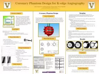
This project aims to design a coronary phantom for K-edge angiography, using monochromatic tunable X-ray to reduce radiation dose and contrast agent concentrations. The goal is to determine the optimal angle for stereo imaging and test contrast agent dilutions.

E N D
Coronary Phantom Design for K-edge Angiography John Jorgensen Sarah Pachtman Punam Patel Marcus Spallek Advisor: Dr. Frank Carroll
MXI Systems • MX 200 • Applications • Radiotherapy • Crystallography • Mammography • Pediatrics • K-edge Imaging • Angiography??? http://www.onlinetelemedicine.com/html/product/sam_images/Angiography.jpg
Project Background • Monochromatic tunable X-ray: • Reduced radiation dose to patient • Reduced contrast agent concentrations • K-edge imaging Diagram of MX200’s laser paths and major elements. http://www.mxisystems.com/mx200.html FOR MORE INFO... Free Electron Laser Laboratory http://www.vanderbilt.edu/fel/
Project Goals • Design acoronary phantom • Test contrast agent concentrations • hypersensitivity reaction to iodine (3-12% patients) • current clinical dilution is 1:16 • using monochromatic plain films it is possible to see 1:256 dilutions • Determine optimal angle for stereo imaging Tan θ = 3/30 = 0.10 θ ~ 6° 3” θ 30”
Solution Description • Radio Translucent at 35 keV • Flexible material • Fluid perfusion • Phantom Size • 7.5cm diameter • Vasculature • Size Range 5 - 0.5 mm • Coronary arteries on surface • Curved channels • Hollow vessels Finger imaged at 19 keV Finger imaged at 26 keV
Phantom Designs SolidWorks Model Shelley Medical Imaging Technologieswww.simutech.com Preliminary Sketch
Physical Properties Flexible cured product 2-component liquid resin system Room-temperature cure 20 Shore A durometer 60 pli tear strength 200 psi tensile strength Elongation at Break 1000% Material - ReoFlex ™ Rubber www.smooth-on.com
November December January February March April Schedule Research Material Testing & Design Manufacturing Testing FOR MORE INFO... http://vubme.vuse.vanderbilt.edu/srdesign/2003/group12/NCIIA%20Grant%20Proposal.htm
Current Status • Work Completed • Material Selected • SolidWorks Design Completed • ZCORP Contacted (Manufacturer) • Rapid Prototyping Machine
Future Plans • March • Send for Prototype • Start Testing • Test Dosimetry for Imaging • Test for Optimal Stereo Angle • Start Final Report
Team and Resources • Advisor: Dr. Frank Carroll, M.D. • Monochromatic x-ray machine • W.M. Keck Free-electron Laser (FEL) Center at Vanderbilt University • Materials and tools provided by BME Department and FEL Center • Polyurethane resins from outside contractors Photograph of device with laser cover removed http://www.vanderbilt.edu/fel/Xray/manuscripts/final%20report%20for%20ONR%20grant%2020021218.doc
Questions? Home