Download
1 / 60
620 likes | 965 Views
HEART BLOCKS AND CARDIAC PACEMAKERS. Arun Abbi Jason Mitchell Jan 21, 2010. OUTLINE. SINUS NODE DYSFUNCTION ATRIOVENTRICULAR BLOCKS INTRODUCTION TO CARDIAC PACEMAKERS INSERTION OF TRANSVENOUS CARDIAC PACEMAKER. HEART BLOCK. RELEVENT ANATOMY

E N D
HEART BLOCKS AND CARDIAC PACEMAKERS Arun Abbi Jason Mitchell Jan 21, 2010
OUTLINE • SINUS NODE DYSFUNCTION • ATRIOVENTRICULAR BLOCKS • INTRODUCTION TO CARDIAC PACEMAKERS • INSERTION OF TRANSVENOUS CARDIAC PACEMAKER
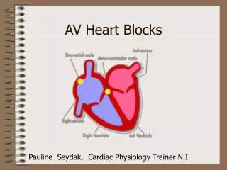
HEART BLOCK • RELEVENT ANATOMY • Conduction: SA > Atrium > AV Node > His > Purkinje Network • AV node highly innervated • Responsive to sympathetic and vagal stimuli • RCA blood supply • His bundle less responsive • Dual blood supply
SINUS NODE DYSFUNCTION • Abnormal sinus impulse formation and propagation • AKA Sick Sinus Syndrome • Umbrella term for: • Sinus bradycardia • Sinus arrest • Sinoatrial exit block • Tachy-brady syndrome
SINUS NODE DYSFUNCTION • ETIOLOGY • Unclear • Fibrosis (most common) • Structural heart disease • Medications • Electrolyte imbalances (HypoK, HypoCa) • Endocrine (HypoTSH, HypoT)
SINUS NODE DYSFUNCTION • SINUS ARREST • Absent sinus P waves > 2 – 3 seconds • Result of absent sinus impulse formation • Duration of pause is not a function of the P-P interval
SINUS NODE DYSFUNCTION • SINOATRIAL EXIT BLOCK • Conduction delay between sinus node and atrium • Three types
SINUS NODE DYSFUNCTION • SINOATRIAL EXIT BLOCK • First Degree • Conduction delay between sinus node and atria • Cannot be identified on ECG • ?Clinical significance
SINUS NODE DYSFUNCTION • SINOATRIAL EXIT BLOCK • Second Degree • Intermittant conduction block • Type I (Wenkebach) – Progressive shortening of P-P intervals – Pause duration less than twice the P interval – Grouped P waves
SINUS NODE DYSFUNCTION • SINOATRIAL EXIT BLOCK • Type II – Pause duration that is a multiple of the P-P interval
SINUS NODE DYSFUNCTION • SINOATRIAL EXIT BLOCK • Third Degree • Complete conduction block from sinus node to atrium • Cannot be distinguished from sinus arrest on ECG • Typically results in an escape rhythm
SINUS NODE DYSFUNCTION • TACHY-BRADY SYNDROME • Bradycardia alternating with brief episodes of SVT • Usually Afib • ???Cause
ATRIOVENTRICULAR BLOCK • ETIOLOGY • Congenital • Acquired – Extensive DDX • Medications • Hyperkalemia (>6.3 mEq/L) • Hypoxia • Increased vagal tone • Ischemia/Infarction (~40%) • Fibrosis (~50%) • Infection/Inflammation • Vascular Disease • Idiopathic • Usually never identified
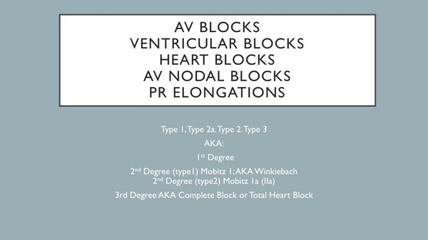
ATRIOVENTRICULAR BLOCK • FIRST DEGREE AV BLOCK • Prolongation of PR > 200 ms • Location of block • AV node, His bundle, His-Purkinje system • Correlate with QRS complex • Prognosis • Framingham: More likely to develop Afib, require permanent pacemaker, and increased all-cause mortality • Locate source of block • If AV node, generally benign and no further Ix • If infranodal, may require His-bundle electrocardiogram • No specific intervention required for stable 1st degree block
ATRIOVENTRICULAR BLOCK • FIRST DEGREE AV BLOCK
ATRIOVENTRICULAR BLOCK • SECOND DEGREE AV BLOCK • Type I (Wenckebach/Mobitz I) - Normal • Gradual prolongation of the PR interval followed by dropped QRS • Atrial impulses reach AV node while it is partially refractory • Location usually the AV node
ATRIOVENTRICULAR BLOCK • SECOND DEGREE AV BLOCK • Type II – Never normal • PR interval constant • Usually a result of underlying structural disease • Location typically His-Purkinje system • High Grade Second Degree • 2 or more consecutively blocked P waves
ATRIOVENTRICULAR BLOCK • SECOND DEGREE BLOCK • Different sites of involvement/prognoses • Type I: Generally involves AV node and is benign • Type II: Almost always infranodal and may progress to 3rd degree (slow unreliable escape) • Difficult to distinguish type in 2:1 conduction block
ATRIOVENTRICULAR BLOCK • THIRD DEGREE BLOCK • Complete AV node failure to conduct • Block may be anywhere in conduction system • Constant P-P and R-R intervals but no relationship • Variable PR intervals, Atrial HR > Ventricular HR • May be hemodynamically unstable • Slow heart rate may produce Torsade , especially in women
HEART BLOCK • ECG PRACTICE
HEART BLOCK • INITIAL ASSESSMENT • Hemodynamic Instability • Fatigue, Dizziness, NV, Diaphoresis • Hypotension • Syncope • Dyspnea • Chest Pain • ACLS Guidelines for Symptomatic Bradycardia • Medications • Β- Blockers • Ca2+ Channel Blockers • Digitalis • Amiodarone
HEART BLOCK • INITIAL ASSESSMENT • Investigations • Stabilize first! • ECG • Bloodwork • Electrolytes • Dig level • Troponin
HEART BLOCK • MANAGEMENT • O2, IV, Monitors • Transcutaneous pacing • Transvenous pacing • > 30 minutes transcutaneous pacing • Unable to obtain capture • Consider atropine • Consider catecholamines (be cautious)
HEART BLOCK • CARDIOLOGY CONSULTATION • Outpatient • New, asymptomatic Type I 2nd Degree (while awake) • Inpatient • Any symptomatic block • New, asymptomatic Type II 2nd Degree • Asymptomatic 3rd Degree • Concomitant MI/Ischemic symptoms • High Grade AV Block
CARDIAC PACING • INDICATIONS • Temporary • Any symptomatic AV block • Asymptomatic, but associated with Torsade • Permanent • ACC/AHA/HRS 2008 Guidelines: • Divided into Class Based Recommendations
CARDIAC PACING • INDICATIONS AV Block • Class I • 2nd and 3rd Degree • Bradycardia with symptoms (C) • Associated arrhythmias and medications that produce symptomatic bradycardia (C) • Asymptomatic, but asystole >3 sec or escape < 40 bpm or wide QRS escape or Afib and bradycardia with systole >5 seconds (C) • After ablation of AV node or unresolving post-op block (C) • Associated with MD, Kearns-Sayre syndrome, Erb dystrophy (B) • Associated with exercise w/o MI (B)
CARDIAC PACING • INDICATIONS AV Block • Class IIa • Asymptomatic persistent 3rd degree with escape > 40 (C) • Asymptomatic 2nd degree with intra or infra-Hisian block (B) • Symptomatic 1st or 2nd degree block (B) • Asymptomatic 2nd degree block with narrow QRS (B) • Class IIb • 1st or 2nd degree with MD, Erb dystrophy, peroneal muscular atrophy +/- symptoms (B) • AV block in setting of drug toxicity when block expected to recur (B)
CARDIAC PACING • INDICATIONS AV Block • Class III • Not indicated for asymptomatic 1st Degree (B) • Not indicated for asymptomatic Mobitz I with block at AV node (C) • Not indicated for AV block that is expected to resolve and unlikely to recur (drug toxicity, Lyme disease, transient increased vagal tone) (B) • Also not indicated in: • PEA Arrest • Traumatic cardiac arrest
CARDIAC PACING • PACING MODES • 5 Position Nomenclature • First 3 Positions most common in pacemaker description • Position I: Chamber being paced • Atrium (A), Ventricle (V), Both (D), None (O) • Position II: Chamber being sensed • Atrium (A), Ventricle (V), Both (D), None (O) • Position III: Pacemaker’s response to sensing • Triggers (T), Inhibits (I), Both (D), None (O)
CARDIAC PACING • PACING MODES • Position IV: Programmability and Rate Control • Hierarchical • Rate Modulation (R), Communicating (C), Programmable (P), (O) • Position V: Antitachydysrrhythmia Function • Pacing (P), Shocking (S), Both (D)
CARDIAC PACING • PACING MODES • Most pacemakers encountered are: • AAIR – Useful for sinus node dysfunction with intact AV node • VVIR – Useful for chronically ineffective atria (AF, AFlutter) • DDD – Most common. Preserves AV synchrony • Reduces risk of AF, reduces signs/symptoms HF, improves QOL • No significant mortality benefit over single-chamber pacing
CARDIAC PACING • ECG MANIFESTATIONS • Depends on Pacing Mode • Atrial Pacing • Small pacemaker spike prior to P wave with normal morphology • Ventricular Pacing • LBBB-like and prolonged, inverted QRS (V5/6) and LAD