Download
1 / 18
190 likes | 247 Views
Explore the intricate details of tooth structure, innervation, and function. Learn about enamel, dentine, pulp, cementum, and more. Delve into the root canal system, nerve fibers, and pain receptors within the dental pulp. Gain insights into the complexity of tooth innervation by trigeminal and facial nerves.

E N D
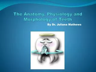
The Anatomy, Physiology and Morphology of Teeth By Dr. Juliana Mathews
Tooth Anatomy • The crown: • consists of enamel, dentine and pulp • The root: • has a root canal with blood vessels and nerves • covered by cementum and held together by periodontal fibres • embedded in the alveolar bone • Enamel: • white hard covering over the crown of the tooth • no nerve or blood supply • cannot heal or repair like bone or dentine • Dentine: • covered by enamel on the crown and cementum on the roots • protects the pulp
Tooth Anatomy continued • Pulp: • Consists of nerves, blood vessels and connective tissue • Found in pulp chamber and root canal • Anastomoses between venules and arterioles • Cementum: • Covers the dentine of the root • Attached to the periodontal ligament • No nerve supply
Tooth Anatomy continued • Periodontium: • Alveolar process: bony extensions of the maxilla and mandible that support teeth • Cortical Plate: dense outer layer of bone covering the spongy (cancellous) bone • Periodontal ligament : • Periodontal fibres attach the roots to the alveolar bone • has a nerve and blood supply • provides an elastic cushion between the tooth and bone • Gingiva: covers the teeth and the alveolar process
Enamel Periodontal Ligaments Dentine Dentinal Tubules Cementum Pulp Alveolar Process Cortical Plate Spongy Bone
Root Canal System • Pulp chamber is found on the coronal part of the tooth • Reduces in size with age due to secondary dentine due to physiological or pathological reasons • Orifices to the root canal are found on the floor of the pulp chamber • Canals taper towards the apex • The narrowest part of the canal is found at the apical constriction, which opens out as the apical foramen and exists to one side i.e. 0.5mm-1mm from the anatomical apex • New layers of cementum are constantly being laid down, therefore the centre of the foramina deviates from the apical centre • Lateral canals can develop between the main body of the root canal and the periodontal ligament space • Accessory canals can develop in the apical region forming the apical delta • Lateral and Accessory canals develop due to a break in the “Hertwigs” epithelial root sheath or during the development, the sheath grows around the existing blood vessel • Lateral canals can be impossible to instrument and can compromise obturation
Root canal system continued • Some roots can have more than one canal and they don’t always merge • Single rooted teeth that have a single canal can end in a single foramen. Some have an apical delta and have a single canal but many exits • Multi- rooted teeth commonly have multiple foramina and each root can have two or three canals. Some canals merge before their exit and some can leave the root independently • Eg. Some maxillary second premolars can have two roots (usually are single rooted) or a single root with 2 canals • Eg. The mesio-buccal root of the maxillary first molar can have two canals (usually one canal present)
Physiology of the Dental Pulp Nerve fibres: • consist of sensory (afferent) fibres, sympathetic fibres and parasympathetic fibres • sensory fibres pass through the apical foramen and end at the peripheral pulp • sensory nerve fibres originate from the trigeminal ganglion • C –fibres: • Unmyelinated, high threshold fibres responding to mechanical, thermal or chemical stimulation • Dull, poor localized pain • A- delta fibres: • myelinated, low threshold mechano- receptors • sharp localized pain • A-beta fibres
Pain • Inflammation of pulp develops: • Increased pulpal pressure against the sensory nerve endings • Sensitized nerves release neuropeptides and cause inflammation= Neurogenic inflammation • A-delta fibres respond to hydrodynamic stimuli • C-fibres respond to the inflammatory mediators • Pheripheral sensory nerves produce pain = hyperalgesia • Peripheral sensory nerves sprout/branch in the inflammed area but disappear as the inflammation subside • Central sensitization occurs when there is a flow of continuous pain impulses which can occur in acute and chronic states
The Innervation of Teeth • Trigeminal Nerve: (CN V) • Three sensory branches • Opthalmic branch supplies the orbit and forehead • Maxillary branch supplies the maxillary sinus and upper jaw teeth • Mandibular branch supplies the tongue and the lower jaw teeth • Facial Nerve: (CN VII) • Motor and sensory branches • Innervates • muscles of facial expression • taste buds of the anterior 2/3 of the tongue • salivary glands
Innervation of Teeth continued • Maxillary Teeth: • Anterior superior alveolar nerve: upper incisors and canines (CNV2) • Middle superior alveolar nerve: upper premolars and the mesio-buccal root of the maxillary first molar (CNV2) • Posterior superior alveolar nerve: upper molars except the mesio-buccal root of the maxillary first molar (CNV2) • Mandibular Teeth: • Inferior alveolar nerve: mandibular teeth, gingiva and lower lip unilaterally (CNV3) • Lingual nerve: anterior 2/3 of tongue and mucosa of the floor of the mouth (CNV3) • Buccal nerve: gingiva on the buccal side of posterior teeth (CNV3)
Blood supply • Maxillary teeth: • Superior alveolar artery: anterior, middle and posterior branch (Maxillary Artery) • Mandibular teeth: • Inferior alveolar artery (Maxillary Artery)
Tooth Morphology • Please look the additional notes for this section