Download
1 / 37
380 likes | 613 Views
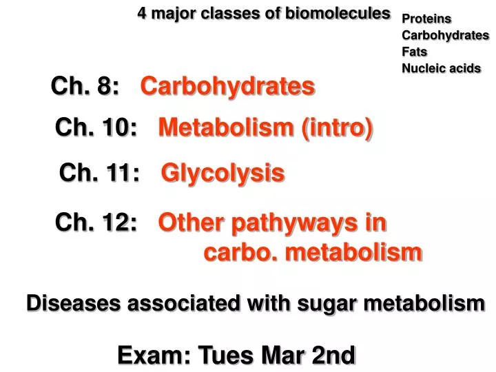
4 major classes of biomolecules. Proteins. Carbohydrates. Fats. Nucleic acids. Ch. 8: Carbohydrates. Ch. 10: Metabolism (intro). Ch. 11: Glycolysis. Ch. 12: Other pathyways in carbo. metabolism. Diseases associated with sugar metabolism. Exam: Tues Mar 2nd.

E N D
4 major classes of biomolecules Proteins Carbohydrates Fats Nucleic acids Ch. 8: Carbohydrates Ch. 10: Metabolism (intro) Ch. 11: Glycolysis Ch. 12: Other pathyways in carbo. metabolism Diseases associated with sugar metabolism Exam: Tues Mar 2nd
Ch. 8: Carbohydrates Most abundant class of macromolecules on the earth Glucose
‘Hydrate of carbon’ Carbohydrate (a.k.a. sugars, saccharide) • (CH2O)n n>3 • Monosaccharide – smallest unit or ‘building blocks’ • Oligosaccharide - disaccharide • Polysaccharide – (more than 20) Glycoconjugates - linked to protein or lipid (2-20) Function Energy storage and release Cell wall and protective coatings Marker mol. on cell surface cell-cell interactions virus invasion… Protein function (covalent modification) DNA/RNA
Polyhydroxyl aldehyde (aldose) or Polyhydroxyl ketone (ketose) Aldotriose Glyceraldehyde (D or L) Ketotriose Dyhydroxyacetone D enantiomer predominate in nature Monosaccharides (CH2O)3
Monosaccharides - Aldoses # Isomers = 2n where n = # of chiral carbons Enantiomer Distant chiral C From most oxidized Epimers – differ in configuration at only one chiral carbon Not all made in nature
Monosaccharides - Ketoses # Isomers = 2n where n = # of chiral carbons
Furanose – 5 membered ring, one member O of –OH Pyranose – 6 membered ring, one member O of –OH Cyclization - Ring Structures Optical behavior of monosaccharides in solution suggests that they have an additional chiral center. Similar to:
Cyclization of Monosaccharides Most oxidized C New chiral C
Cyclization - aldohexose • Draw most oxidized carbon (C1 aldose and C2 ketose) on right and number C clockwise • In ring most oxidizes carbon new chiral center (anomeric C) • Transfer information from Fisher projections • -OH on right then down in Haworth • -OH on left then up in Haworth • Bulky substituent on highest numbered carbon points up Anomers rapid equilibrium
Equilibrate in solution In solution at 31°C 64% b-D-glucopyranose 36% a-D-glucopyranose Very little in open chain or furanose form Cyclization - aldohexose Anomers Anomers
Cyclization - aldopentose Equilibrium Anomeric C Hemiacetal Haworth projection Anomers
“Furanose” Conformations Not planar Rapidly interconvert
“Pyranose” Conformations More stable Whether a ring substituent is Equatorial (same plane) or Axial (above/below) depends on whether C-1 or C-4 is above the ring.
Important in metabolism Derivatives of monosaccharides – sugar phosphates alcohol phosphate esters Energy metabolism hemiacetal phosphate Nucleic acid metabolism More reactive
replacement of one of the -OH groups with H Important in DNA Derivatives of monosaccharides –deoxy sugars
RNA DNA OH OH OH OH
amino groups or an acetylated amino group replaces one of the -OH groups Derivatives of monosaccharides – amino sugars NeuNAc Sialic acids: on cell surface glycoproteins
reduction of carbonyl oxygen, so polyhydroxyl alcohol Derivatives of monosaccharides – sugar alcohols Id glyceraldehyde Idose ---- Inositol
derived from aldoses by either the oxidation of C1 or the highest-numbered carbon Glucose oxidation : gluconate or glucuronate gluconate can cyclize under acidic conditions to form a lactone - intramolecular ester. Derivatives of monosaccharides – sugar acids
Primates unable to do this reaction Vitamin C or L-Ascorbic Acid
Glycoside Bonds – • acetal linkage between the anomeric carbon of a sugar and an alcohol, an amine, or a thiol • Compounds containing glycoside bonds are called glycosides if glucose donates the anomeric carbon then glucosides
Glycoside Bonds – Disaccharides No open chain equil Hemiacetals -a reactive carbonyl that can be oxidized. reducing non-reducing b anomer: refers to free C1 OH (In equilibruim) non-reducing sugar
Glycoside Bonds – Disaccharides epimer Most abundant disacc. in nature (plants)
Glycoside Bonds – Reducing and Non-reducing • Since mono- and disaccharides are hemiacetals they have a reactive carbonyl that can be oxidized. • Linear polymer usually one reducing end (free anomeric carbon), one non-reducing end, and all internal monosaccharides are acetals that are not in equilibrium with open chains form. • Some polymers such as the disaccharide sucrose do not have a reducing end (both anomeric carbons are involved in the gycosidic bond) so non-reducing sugar.
Glycoside Bonds – Other
Plant starch – mixture of amylose and amylopectin Animals glycogen Polysaccharides –Glucose Storage • Homoglycans- one type of monosaccharide Amylose • 100-1000 glucose residues (maltose units) Amylopectin and Glycogen Amylopectin: branch every 25 residues Glycogen: branch every 8-12 residues 10% mass of liver • No template (ie no gene)
Humans digest starch via two enzymes: α -amylase - endoglycosidase of α-(1-4) linkages (random) debranching enzyme (cleaves limit dextrans) Higher plants have β- amylase exoglycosidase of α- (1-4) linkages, releasing the disaccharide maltose Polysaccharides -Starch Degradation Know how starch is broken down ! Single reducing end
Humans don’t have b-glucosidases Microbe that live in ruminants do Polysaccharides –Structure Amylose • Cellulose • b-(1-4) linkage 180 deg rotation 300- 15,000 Glc residues Rigid extended conformation H-bonding Forms bundles or fibrils Plant cell walls, stems and branches termites
Chitin found in exoskeletons of insects and crustaceans, and in cell wall of algae and fungi composed of β- (1-4)linkage of GlcNAc residues. Polysaccharides –Structure 2nd most abundant organic compound on earth 180 deg rotation H-bonding Adjacent strands
Glycosaminoglycans have dissaccharide components (repeating) one sugar is an amino sugar; e.g. GalNAc, or GlcNAc. The other sugar is usually a uronic acid Certain types can be sulfated, etc. They are highly hydrated, and viscous and are excellent lubricants Glycoconjugates: Proteoglycans unbranched heteroglycan Fluid of joints Elastic and resistant to compression cartilage cartilage
Bacteria cell wall, heteroglycans chains linked to peptides GlcNAc linked to N-acetylmuramic acid (MurNAc) joined by β -(1-4) linkage Glycoconjugates: Peptidoglycan Large/rigid mol Defines shape of cell Gram stain +/-
O-linked - typically a GalNAc residue linked to the side chain of Ser or Thr, occurs in the golgi N-linked-typically a GlcNAc residue linked to the nitrogen of an Asn, occurs in the endoplasmic reticulum Glycoconjugates - Glycoproteins
Glycoconjugates - Glycoproteins N-linked Large amt of structural diversity possible !!
Glycoconjugates - Glycoproteins and blood types
Draw the Fisher projections of fructose and show how it can cyclize to form both the α and β anomers of fructopyranose and fructofuranose. Draw the disaccharide b-D-ribofuranosyl –(1-4)-a-D-glucopyranose. Is this a reducing or nonreducing sugar? Compare and contrast the structures of starch, glycogen and cellulose. Practice Problems