Download
1 / 13
130 likes | 143 Views
A comprehensive thoracic exam including vital signs, appearance, sensory assessment, range of motion, and orthopedic tests. This exam evaluates the thoracic region for any abnormalities or conditions.

E N D
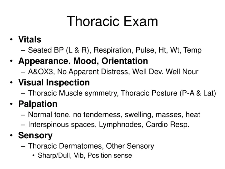
Thoracic Exam • Vitals • Seated BP (L & R), Respiration, Pulse, Ht, Wt, Temp • Appearance. Mood, Orientation • A&OX3, No Apparent Distress, Well Dev. Well Nour • Visual Inspection • Thoracic Muscle symmetry, Thoracic Posture (P-A & Lat) • Palpation • Normal tone, no tenderness, swelling, masses, heat • Interspinous spaces, Lymphnodes, Cardio Resp. • Sensory • Thoracic Dermatomes, Other Sensory • Sharp/Dull, Vib, Position sense
Thoracic Exam ROM • Flexion • 20-40 degrees • Extension • 25-45 degrees • Rotation • 35-50 degrees • Lat Flexion • 20-40 degrees
Seated Spinous Percussion Schepelman’s Chest Expansion Dejerine’s Triad* Valsalva* Thoracic Exam Orthopedics Standing • Gait • Adam’s Sign Supine • Soto-hall • Sternal Compression • Beevor’s Sign * This test shows up in more than 1 Exam
Thoracic Exam Orthopedics • SPINAL PERCUSSION: • Procedure: patient seated with head flexed, dr. percusses the sp’s and paravertebral musculature. • (+): if localized or radiating. • Indicates: 1. local – fracture or severe sprain. • 2. radicular – IVD syndrome. • Clinical note: ultrasound may aggravate the condition.
Thoracic Exam Orthopedics • SCHEPELMAN’S SIGN: • Procedure: patient seated and raises arms and then bends laterally to each side. • (+): if pain on either side. • Indicates: pain on concave side = intercostal neuritis. • Pain on convex side = intercostal myofascitis.
Thoracic Exam Orthopedics • Chest Expansion
Thoracic Exam Orthopedics • DEJERINE’S TRIAD*: • Procedure: patient seated, then cough, sneeze, and strain. • (+): pain. • Indicates: disc protrusion, spinal cord tumor or spinal compression fracture.
Cervical Exam Orthopedics • VALSALVA*: • Procedure: do I have to explain??? • (+): /reproduce pain. • Indicates: space occupying lesion (herniated disc or tumor), or osteophyte.
Thoracic Exam Orthopedics • Gait
Thoracic Exam Orthopedics • Adams Sign
Thoracic Exam Orthopedics • SOTO-HALL: • Procedure: patient supine with hands above head and dr. passively flexes the patient’s chin to chest, while holding onto the sternum. • (+): pain. • Indicates: subluxation, sprain/strain, fracture, disc lesion, exostoses, or meningitis.
Thoracic Exam Orthopedics • STERNAL COMPRESSION: • Procedure: patient supine and dr. presses on sternum. • (+): if pain. • Indicates: localized pain at lateral border of rib = rib fracture.
Thoracic Exam Orthopedics • BEEVOR’S SIGN: • Procedure: patient supine and does a ½ sit up and dr. inspects the umbilicus for deviation. Then patient lies flat and lifts the legs up, while dr. palpates and observes the umbilicus. • (+): if umbilicus deviates in any direction. • Indicates: a spinal cord lesion at T7 – T10 depending on where the umbilical deviation was. • Clinical note: in the presence of prolonged illness followed by lower extremity paresthesia, this test needs to be performed.