Download
1 / 28
370 likes | 541 Views
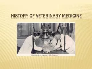
Explore the history of diagnostic medicine from ancient times to present day advancements, including major milestones, key tools, and influential figures. Learn how technology has shaped the future of radiology, medical laboratories, and diagnostic tools.

E N D
Bell Work • What is diagnostics? • Name 3 diagnostic tools? • Where do diagnostic professionals work?
Standard 3: Investigate and document the history of radiology, medical laboratories, and other areas of diagnostic medicine. Explain how technology is influencing the future of each area. • At the end of this standard I CAN: • Identify major advancements in dx medicine and place them in chronological order through use of a timeline • Actively participate in class discussion during lecture to explain how technology influences diagnostic medicine • Select one diagnostic tool and trace the advancement of the tool through group collaboration
What is diagnostics? • adjective • 1. of, relating to, or used in diagnosis. • 2. serving to identify or characterize; being a precise indication. • a device or substance used for the analysis or detection of diseases or other medical conditions. • A medical professional in the field of diagnostic medicine has the assignment to accurately classify symptoms, detect a patient's disease and to find the best available treatment option for the given illness.
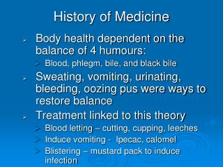
Ancient times • Diseases and illness were thought to be a punishment from the gods • Caused by evil spirits and demons • Used herbs and plants as medicine • Priests acted as physicians and treated sick people in the temple • Make up of the body was unknown • Hippocrates is now knows as the “Father of Medicine” due to his hypothesis that nutrition and cleanliness prevents illness and disease
Middle ages (a.d.800-1400) • Interest in medical practices grew • Medical universities were established in the 9th century • An outbreak of bubonic plague in the 1300s resulted in the death of 75% of the people in Europe and Asia • Became apparent that diseases are contagious and quarantine laws were enacted
The Renaissance (a.d.1350-1650) • The “Rebirth of the science of medicine” • Human dissection to view body organs • Artists took strong interest • Medical thermometer devised by Italian physician Sanctorius • First diagnostic tool
16th, 17th, 18th Centuries • Knowledge regarding the human body greatly increased • Microscope developed by Janssen • Microscope improved upon by Leeuwenhoek • Followed by Hooke
19th Century • Rene Laennec invented the stethoscope • Louis Pasteur known as the “father of Microbiology” • Proved microorganisms cause disease • Discovered pasteurization kills bacteria in milk • Robert Koch • Created the culture plate • Identified germ causing TB • Wilhelm Conrad Roentgen discovered X-Rays in 1895.
Video • https://www.youtube.com/watch?v=gsV7SJDDCY4
20th Century • Chest x-ray allowed for early detection of TB
20th Century • 1906: First x-ray contrast medium • 1910: Barium sulfate introduction for GI diagnosis • 1910: Theory of Radioactivity published by Marie Curie • 1912: Investigation of x-ray radiation for patient therapy
20th Century • 1906; Electrocardiograph (ECG/EKG) invented by Willem Einthoven
20th Century • 1929: First cardiac catheterization performed by Forssmann on himself • 1945:Coronary artery imaging allowing visualization of blood vessels that feed the heart
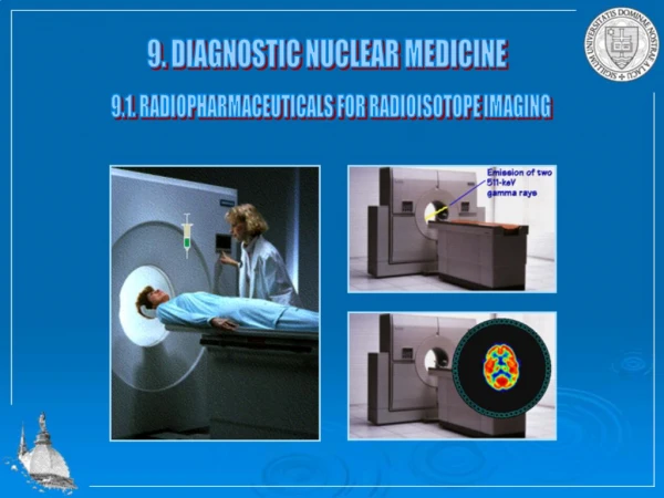
20th century • 1950’s: Blood chemistry tests became readily available • Quickly ID infection V. Virus • Electrolyte imbalances • 1950’s:Nuclear medicine developed making radiation within the patient instead of emitted from machine • 1955: Panoramic x-ray of jaw
20th Century • 1960; Ultrasound imaging developed to look at the abdomen, kidneys, fetal baby, carotid blood vessels, and heart • 1970: X-ray mammography finds widespread application in imaging the breasts
20th Century • 1972: Computed Tomography scanning invented
20th Century • 1976: Coronary angioplasty used • 1980: Magnetic Resonance Image (MRI) first introduced • 1985: Clinical Positron Emission Tomography (PET) scanning developed allowing for 3D images
Present day • CT Angiography allows quick access to view of vessels without invasive procedure • Advanced medical imaging replacing exploratory surgery • Advancement in PET scanning and digital imaging allowing for early Cancer diagnosis • Bedside blood testing equipment allowing for instant treatment • REMEMBER DIAGNOSTICS=DIAGNOSIS
Understanding Timeline • A timeline is a method for picturing or seeing time. Historians take events and place them on a timeline. This shows the chronology of a span of time. By doing this, the relationship between events can be seen. • A KEY TO REMEMBER! If you’re trying to remember dates, thinking in terms of centuries can really be confusing. So, whenever you hear a time period given as a century, always translate the century into years in your head. For example: ■ When you see 14th century, think 1300’s ■ When you see 6th century, think 500’s
Create a Timeline Instructions • Pick out 7 items most interesting and relate to diagnostics in your era. Two of the items need to come from my PowerPoint on Web site. • Visually display numbers (in order)on timeline. Must have a picture to depict each item you choose. Your group will present timeline in class tomorrow after lunch
Create a Timeline Group 1: Ancient Times/Middle Ages Group 2: 17th Century Group 3: 18thCentury Group 4: 19thCentury Group 5: 20thCentury History of medical labs History of radiology History of Optometry History of Cardiology