Download
1 / 60
1.22k likes | 4.17k Views
Radiation Hazards, Effects & Risks Radiation Protection for X-ray Technologists. Dr Tim Wood Clinical Scientist. Overview. A little bit of history The discovery of radiation and health effects A bit of radioactive quackery c1920 The mechanisms of radiation injury

E N D
Radiation Hazards, Effects & RisksRadiation Protection for X-ray Technologists Dr Tim Wood Clinical Scientist
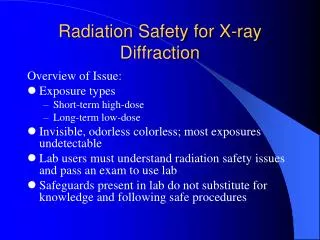
Overview • A little bit of history • The discovery of radiation and health effects • A bit of radioactive quackery c1920 • The mechanisms of radiation injury • The Physics/Biology of radiation induced effects • What is radiation dose? • Deterministic and stochastic effects (with examples – not for the squeamish) • So how big are the risks? • Where do we get our data from? • Putting risk into context • Why do we need to worry about radiation dose in dentistry?
The discovery of radiation • Wilhelm Röntgen discovered X-rays on 8th Nov 1895. • Took first medical X-ray of wife’s hand (22nd Dec 1895). • Used to diagnose Eddie McCarthy’s fractured left wrist on 3rd Feb 1896 (20 min exposure). • Awarded first Nobel Prize in Physics in 1901 for his discovery of ‘Röntgen rays’.
The discovery of radiation • Henri Becquerel discovered radioactivity on 26th Feb 1896.
The discovery of radiation • In 1903 he shared the Nobel Prize for Physics with Pierre and Marie Curie who had refined Becquerel’s work and discovered the existence of Radium.
Then just as quickly… • Late 1896 • Elihu Thomson – First person to prove a direct relation between exposure and health effects. • Deliberately exposed left index finger for half an hour a day for several days – resulted in erythema, swelling and pain.
Then just as quickly… • Late 1896 • Edison’s assistant – hair fell out & scalp became inflamed & ulcerated. • Several reports of ill-effects on the eyes – result of direct viewing of fluorescent screens.
Edmund Kells • April 1896 built own X-ray machine, packed films in rubber and took X-ray of his dental assistant. • 10 years on, cancer of right hand. • 42 operations in next 20 years – lost hand, arm and shoulder.
Sister Blandina (1871-1916) • 1898 – Radiographer in Cologne. • Held nervous patients & children with unprotected hands. • 6 months later had cancer of hand – arm amputated. • 1915 – severe breathing difficulties. • Extensive shadow on the left side of thorax. • Large wound on her whole front- and back-side. • Died 22nd Oct 1916.
Mihran Kassabian (1870-1910) • Was a medical missionary student and photographer who became one of the most prominent pioneering radiologists of the time. • By 1898 he had become an American citizen
The literature c1901… • Rollins W. X-light kills. Boston Med Surg J 1901;144:173. • Codman EA. No practical danger from the x-ray. Boston Med Surg J 1901;174:197
But by 1913… • Dental office. • Lead glass shield around the tube (but none around the high voltage wires!).
Radioactive quackery • 1903 – J. J. Thomson identifies radioactivity (Radon) occurring naturally in well water. • c1910 – Radioactive health springs cured everything (apparently)… • 1912 – The Revigator was launched to provide radioactive water at home. • Many other products followed. • drinks, inhalers, etc.
Radioactive quackery • c1920-1930 – Radium became the alternative to Radon with all manner of products available. • e.g. salves, beauty creams, toothpaste, ear-plugs, chocolate bars, bread, soaps, suppositories and contraceptives.
Mechanisms of Radiation Injury • LD(50/30) = 4 Gy • The dose required to kill 50% of test cohort in 30 days. • 280 J to 70 kg man. • 1 milli-Celsius rise in body temp. • drinking 6 ml of warm tea. i.e. the damage is not caused by heating, but ionisation.
Have the ability to separate electrons from atoms to produce ‘ions’ X-rays Gamma rays (-rays) Beta particles () Electron beams Ionising Radiations
Damage depends on a number of factors: • The type and number of nucleic acid bonds that are broken. • The intensity and type of radiation. • The time between exposures. • The ability of the cell to repair the damage. • The stage of the cell’s reproductive cycle when irradiated.
What is radiation dose? • Absorbed Dose (Jkg-1 or Gy) • Amount of energy deposited per kilogram. • Dose to an organ or tissue. • DOSE TO A CERTAIN PLACE IN THE BODY • Effective Dose (Jkg-1 or Sv) • This is the average dose to the whole body. • This gives us the risk of contracting cancer due to the X-ray exposure. • OVERALL DOSE TO THE WHOLE BODY RADIATION TISSUE
Deterministic effects of Ionising Radiation • Where very large doses kill many cells • Radiation ‘burns’. • Cataract. • Radiation sickness. • Deterministic effects • Caused by significant cell necrosis. • Not seen below a threshold dose. • Will definitely be seen above a threshold dose. • Above the threshold, the bigger the dose, the worse the effect.
Hair loss from CT scan (Imanishi et al 2005) • 53-year-old woman with subarachnoid haemorrhage. • 4 CT perfusion scans and two angiographies of the head performed within first 15 days of admission . • Bandage-shaped hair loss 37 days after first CT lasted for 51 days.
2 embolisations – hair loss after each procedure. 13 x 10 cm area. Re-grew after 4 months. Dose estimate >3 Gy.
Interventional Cardiology • 40 year old male • 4 procedures on one day 29th March 1990 • Coronary angiography • Coronary angioplasty • Second angiography due to complications • Coronary artery by-pass graft
Probably >20 Gy. Extreme case! 6-8 weeks 16-21 weeks 18-21 months Close up of previous Post-skin graft
49-year-old man who underwent two transjugular intrahepatic portosystemic shunt (TIPS) placements and one attempted TIPS placement within a week 6 months – secondary ulceration with surrounding rings of de- and hyperpigmentation. 7.5 months – small blisters developed. Wound is very painful. 10 months – wound has progressed in size and depth. 22 months – non-healing ulcer with exposure of deep tissues, including spinous process of vertebra. 23 months – musculocutaneous skin grafting was performed. Disfigurement is permanent. Koenig, T. R. et al. Am. J. Roentgenol. 2001;177:3-11
Stochastic Effects • Caused by cell mutation leading to cancer or hereditary disease. • Current theory says, no threshold . • The bigger the dose, the more likely effect (but no more severe).
Cancer risks • It is assumed that any dose of radiation could potentially cause cancer. • i.e. a bit like crossing the road – the more times you cross the more likely you are to be run over, but probably never will.
Attributable lifetime risk of fatal cancerdepending on age at exposure Attributable lifetime risk, % per sievert Age at time of exposure
Radiation Effects • Acute radiation syndrome • Including vomiting, diarrhoea, reduction in the number of blood cells, bleeding, epilation (hair loss), temporary sterility in males, and lens opacity (clouding) • Late 1940’s Dr Takuso Yamawaki noted an increase in leukaemia • 20% of radiation cancers were leukaemia (normal incidence 4%) • Incidence peaked at 6-8 years • Solid cancers – excess seen from 10 years onwards.
Cancer deaths between 1950 and 1990 among Life Span Study survivors with significant exposure (i.e. > 5 mSv or within 2.5 km of the hypocentre)
Data Sources for Risk Estimates • North American patients - breast, thyroid, skin • German patients with Ra-224 - bone • Euro. Patients with Thorotrast - liver • Oxford study - in utero induced cancer • Atomic bomb survivors - leukaemia, lung, colon, stomach, remainder.
Hereditary Effects • Observed in animal experiments. • Not observed in A-bomb victims. • ICRP Detriment for severe hereditary disease = 1.3 x 10⁻⁵ [1 in 77000] per mSv. • (i.e. approx 1/4 fatal cancer risk).
Risks associated with X-rays • Adult Exposure (per 1 mSv) • Fatal cancer (all types) 1 in 20,000 • Fatal leukaemia 1 in 200,000 • Non fatal cancer 1 in 100,000 • Heritable effects 1 in 80,000 • Childhood exposure • Fatal cancer 1 in 10,000 • Foetal exposure • Fatal cancer to 15 years 1 in 33,000 • All cancers to 15 years 1 in 17,000 • Heritable effects 1 in 42,000
Average Risk of Death from Various Accidents (US) AccidentType Total Number Individual ChancePer Year Motor Vehicle FallsFires and Hot SubstancesDrowning FirearmsAir TravelFalling ObjectsElectrocutionLightning TornadoesHurricanesAll Accidents 55,791 17,8277,4516,1812,3091,7781,271 1,1481609193111,992 1 in 4,000 (1 in 20000 UK) 1 in 10,0001 in 25,0001 in 30,0001 in 100,0001 in 100,0001 in 160,0001 in 160,0001 in 2,000,0001 in 2,500,0001 in 2,500,0001 in 1,600
30 January 2004 700 CANCER CASES CAUSED BY X-RAYS X-RAYS used in everyday detection of diseases and broken bones are responsible for about 700 cases of cancer a year, according to the most detailed study to date. The research showed that 0.6 per cent of the 124,000 patients found to have cancer each year can attribute the disease to X-ray exposure. Diagnostic X-rays, which are used in conventional radiography and imaging techniques such as CT scans, are the largest man-made source of radiation exposure to the general population. Although such X-rays provide great benefits, it is generally accepted that their use is associated with very small increases in cancer risk. Average X-ray examination dose = 0.5mSv ► 1 in 40,000 risk UK Radiology = 41.5 million X-rays per year
Largest exposure from man-made radiation is medical >41.5 million medical & dental x-raysin UK annually 49
Everyday non-occupational exposure Effective dose from natural background radiation in the UK is approximately 2.3 mSv This natural radiation comes from cosmic rays, rocks and soil, food, human body & radon.