Download
1 / 36
360 likes | 374 Views
This article discusses the changes in DNA (mutations) and their effects on protein structure. It covers topics such as single nucleotide insertions/deletions, reading frame concept, point mutations, larger scale mutations, and alterations of protein structure. It also explores the functions of proteins and the use of PCR and DNA sequencing techniques. Additionally, it touches upon the second messenger systems and their role in signal transduction.

E N D
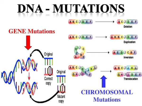
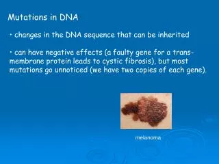
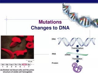
Changes in DNA (Mutations) • Single nucleotide insertions/deletions • Concept of a reading frame • Effect of insertions/deletions depend in part on whether they result in a frame shift • Single alterations “point mutations • Some are completely silent – no change in protein sequence • Larger scale mutations • Generation of a signal for the translation machinery • stop or start codon • splice site. • Changes are heritable and their emergence as a phenotype depends on the effect of this mutation
Protein structure • Proteins are made from amino acids (AAs) – similar sequences of AAs lead to similar protein structure and function. These sequences are referred to as primary structure • Primary structure is the most elementary determinant of protein shape and also is critical for determining sites where proteins are cleaved by various enzymes (proteases). • Proteins adopt specific shapes through a process known as folding. Folding gives rise to secondary and tertiary structure and is primarily determined by the size and charge of the side chains on the amino acids making up the primary structure
Secondary structure • Some proteins have segments that adopt highly sterotyped structures, the most common of these are the the alpha helix and the beta sheet. • These structural elements are extremely thermodynamically stable and can arise from many different primary sequences.
Tertiary structure • The complete 3D structure of a protein • Determined by side chain interactions • Disulfide bonds – very strong • Hydrogen bonds • Electrostatic interactions • Coordination with other atoms – e.g. metal ions like iron
Quaternary structure • Interactions of multiple tertiary structures to generate a functional prtoein
Alterations of protein structure • Transmembrane proteins • Glycosylation • Phosphorylation – • a big one • reversible, specific changes in protein function • Proteolysis
Functions of proteins • Structural proteins • Enzymes • Transport proteins
http://telstar.ote.cmu.edu/biology/animation/biochemistry/SerineProtease/index.htmlhttp://telstar.ote.cmu.edu/biology/animation/biochemistry/SerineProtease/index.html
http://telstar.ote.cmu.edu/biology/animation/biochemistry/SerineProtease/index.htmlhttp://telstar.ote.cmu.edu/biology/animation/biochemistry/SerineProtease/index.html
PCR requires a temperature-stable DNA polymerase • From the geysers of Yellowstone we got Taq polymerase Thermus aquaticus bacteria
How it works • Once a DNA target has been chosen, there are several rules of thumb for primer design that are important to consider. These general rules are:Primer length: Choose primers that will anneal to complementary sequences that are 18-24 nucleotides long.Duplex stability: Both primers in a PCR reaction should have similar melting temperatures (Tm) to ensure that they will have the same hybridization kinetics during the template annealing phase.Non-complementary primer pairs: The two primers cannot share complementarity at the 3’ ends or else they will give rise to primer dimer products.No hairpin loops: Each primer needs to be devoid of palindromic sequences which can give rise to stable intrastrand structures that limit primer annealing to the template DNA.Optimal distance between primers: This is very application specific, but for most diagnostic PCR assays, it is best when the opposing primers are spaced 150-500 bp apart. http://www.biochem.arizona.edu/classes/bioc471/pages/Lecture12/Lecture12.html
Enzyme kinetics where, k1 is the forward rate constant for substrate binding k-1 is the reverse rate constant for substrate binding. k2 is the catalytic rate constant (containing terms related to the transition state).The ES complex is also called the "Michaelis complex".
Also RT-PCR • PCR from RNA
What is it used for? • Amplifying DNA in a sequence-specific manner • Detecting DNA sequences
DNA sequencing http://seqcore.brcf.med.umich.edu/doc/educ/dnapr/sequencing.html
Second messenger systemssignal transduction • What are they? • What are they good for? • Examples • Calcium/CamKII • PKA • How can we interfere with them?
What are they? • First messenger = the neurotransmitter • The binding of the first messenger causes release of a second messenger which diffuses within or between cells • Three broad classes of second messenger systems • G-protein coupled receptors • Ions (calcium) • Tyrosine kinase receptors • The second messenger then goes and does something – activates an enzyme, binds to a channel, etc, leading to a change, e.g. phosphorylation.
What good are they? • Prolonged responses • Long-lasting effects • Multiple inputs can be integrated across long time periods • Highly sensitive responses • Phototransduction or pheromone transduction • Modulatable responses • Response may depend on state of cell or activation of other second messenger systems • Responses that are not direct effects of voltage changes • Changes in input/output curves, changes in cell excitability • May be global changes e.g. via protein synthesis
What activates them? • Activity (via e.g. calcium) • Neurotransmitters/modulators • ACh • Glutamate • GABA • Dopamine • NE • Sensory stimuli • Light • Smells/tastes
What receives the message? • Channels • Open/close (e.g. cyclonucleotide gated channels) • Change their voltage-dependence of activation or inactivation • Examples include modulation of neuronal excitability (postsynaptic), neurotransmitter release (poresynaptic) • Transcription factors • Alter gene expression • Kinases/Phosphatases • Cause phosphorylation/dephosphorylation of other proteins,including the above.