Download
1 / 14
140 likes | 162 Views
This study provides an overview of the Factor V Leiden assay, explaining the mutation's pathophysiology and epidemiology. It evaluates the assay's efficacy, highlighting its importance in assessing thrombosis risk. The extraction, amplification, and genotyping processes are detailed along with the limitations and benefits of this diagnostic tool.

E N D
Factor V Leiden Detection and Genotyping August 6, 2008 Marylin Bicak
Objective • 1. To provide an overview of the Factor V Leiden assay • 2. To explain the pathophysiology and epidemiology of the Factor V Leiden mutation. • 3. To evaluate the efficacy of the assay.
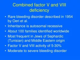
Pathophysiology • inherited condition • autosomal dominant • chromosome 1, gene F5 • Single point mutation, G1691A (arginine to glutamine at 506th amino acid • mutation prevents inactivation of factor V in clotting process (resistant to inactivation by Protein C) • overproduction of thrombin= excess fibrin formation • excess clotting (DVT and PE)
Epidemiology • Most common inherited coagulation disorder in U.S. • 5% Caucasians • 2% Hispanics • 1.2% African Americans • <0.5% Asian Americans • Heterozygous= 3-8 fold increase risk of thrombosis • Homozygous= 30-140 increase risk of thrombosis • risk factors • treatment
Factor V Leiden Assay • 1. Extraction- MagNA Pure Compact Nucleic Acid Isolation Kit and instrument (Roche Diagnostics) • 2. Amplification/Detection/Genotyping- Factor V Leiden Kit and LightCycler 2.0 instrument (Roche Diagnostics)
Extraction • Specimen- EDTA whole blood (200 ul whole blood= 100 ul purified product) • MagNA Pure Kit- pre-sealed reagent cartridge; disposable pipette tip tray assembly; sample tube; elution tube • MagNA Pure Compact Instrument-automated method based on magnetic glass particle technology
Principle of Magnetic Glass Particle Technology • Four step process: 1-lysis; 2-bind to magnetic particles; 3-wash; 4-elution
Amplification-real-time PCR • target- 222 bp fragment of Factor V gene • Factor V Leiden kit- master mix reagents (primers and probes) • LightCycler 2.0 instrument- rapid- 45 cycles (30 min) • Cycle temperatures- denaturation 95C; annealing 60C; elongation 72C • thermal cycler- heat/ambient air cycle • reaction vessel- 20 ul glass capillary
Detection • FRET (fluorescence resonance energy transfer)
Genotyping • Melting Curve Analysis
Summary • Limitations- technical process/ physical space/ instrumentation • Erroneous results- a. false positive (3 rare mutations span same mutation probe) and b. patient sample with elevated WBC • Overall, innovative system and important diagnostic tool for clinical lab