Download
1 / 43
450 likes | 638 Views
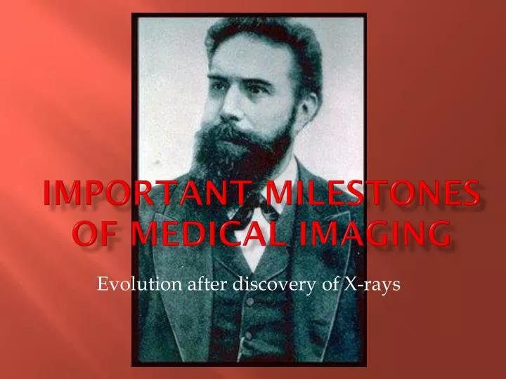
Important milestones of medical imaging. Evolution after discovery of X-rays. X-rays discovered by German physicist Wilhelm Conrad Roentgen. He also produced the first x-ray picture of the body (his wife's hand) in 1895. . 1895.

E N D
Important milestones of medical imaging Evolution after discovery of X-rays
X-raysdiscovered by German physicist Wilhelm Conrad Roentgen. He also produced the first x-ray picture of the body (his wife's hand) in 1895. 1895
Chest x-raywidespread use of the chest x-ray made early detection of tuberculosis (which was the most common cause of death) 1900
X-ray IV contrast mediumFirst contrast filled image of the renal system (kidneys). 1906
Barium sulfateIntroduction as contrast agent for gastro-intestinal diagnosis. 1910
Theory of Radioactivity published by Marie Curie and investigation of radiation for patient therapy(e.g. treatment of cancer). 1910/1912
Radiographic imaging of the gallbladder, bile duct and blood vessels for the first time. 1924
Sri LankaStart of Radiological services in Sri LankaDr. H.O.Goonawardane – first Radiologist 1925
First trained Radiographers in Sri Lanka appointedMr. M.L.B.J. Caspersz & Mr. J.A.N. Fernandopulle 1928
Cardiac catheterizationfirst performed by Forssmann on himself. 1929
Publication“Positioning in radiography” by K.C.Clark - UK 1939
Coronary artery imagingVisualization of (blood vessels that feed the heart). 1945
In Sri LankaFounded the Government X-ray Technical Officers’ AssociationPublication of the journal “Ceylon Radiographer” 1948
Publication“Atlas of Radiographic positions” by Vinita Merill - America 1949
Nuclear Medicine applied in imaging the kidneys, heart, and skeletal system. 1950
X-ray Image Intensifier-Television unitsTo allow dynamic x-ray imaging of moving structures. 1955
Inauguration of Sri Lanka School of Radiography by Mr. Jaundrell Thompson B.Sc., F.S.R 1957
Ultrasound imagingusing high frequency sound waves to look at the abdomen and kidneys, fetal baby, carotid blood vessels and heart. 1960
X-ray mammographyfinds widespread application in imaging the breasts. 1970
Computed Tomography (CT) scanning invented by British engineer Godfrey Hounsfield of EMI Laboratories, England, and South African born physicist Allan Cormack of Tufts University, Massachusetts. 1972
In Sri LankaFounded the Society of Radiographers – Sri Lanka 1975
Magnetic Resonance Imaging (MRI) of the brain was first done on a clinical patient. MRI was developed by Paul Lauterbur and scientists at Thorn-EMI Laboratories, England, and Nottingham University, England. 1980
Radiography training around the world changed from 2- year diplomas to 3-yearhigher diplomas and then to 3-4 year bachelors' degrees
3-Dimensional image processing using digital computers and CT or MR data, three dimensional images of bones and organs were first made. 1984
Positron Emission Tomography (PET)Clinical scanning developed by scientists at the University of California 1985
PACSClinical Networks were first implemented to allow digital diagnostic images to be shared between physicians via computer network, allowing a doctor in Boston to review a CT examination from a patient in Beijing, China 1985
Spiral CTallows fast volume scanning of an entire organ during a single, short patient breath hold of 20 to 30 seconds. Spiral CT had caused a renaissance in CT and lead the way to significant developments like CT Angiography. 1989
MR Angiographydeveloped and clinically available to allow non-invasive imaging of the blood vessels without radiation or contrast injection. 1989
Echo Planar MR Imaging (EPI)Developed and clinically available to allow MR systems to provide early detection of acute stroke. EPI also makes possible functional imaging, for instance of brain activity allowing doctors to investigate the function of different centers of the mind. 1993
Open MRIDeveloped to allow MR scanning of severely claustrophobic or obese patients who could not tolerate conventional MR imaging in a close bore system. 1993
PET/CT scannerInvented by Dr. Ron Nutt and Dr. David Townsend The first generation of PET/CT scanners included a single slice spiral CT integrated with a PET camera which utilized BGO detectors.SPECT/CT also was introduced 2000
Radiotherapy Many advancements have taken place. (This will be dealt with in the lecture by a Medical Physicist today)
In Sri LankaInauguration of First BSc. Radiography programme at University of Peradeniya 2005
In sri Lankafirst ever BSc. graduate radiographers pass out 2011
The greatest tribute we can pay to roentgen is to provide a better service to the sick and the needy.UPGRADING THE TECHNOLOGY ALONE CANNOT ACHIEVE THAT UNLESS THE COMPETENCY OF THE USER IS OF HIGH STANDARD
In almost all the countries radiography courses today are University degree courses.This has become necessary to meet the advancement in technology and requirement in accurate diagnosis and therapy.
It is necessary to make avenues to advance our knowledge in keeping with international levels and standards.An external degree programme is an appropriate and practical solution and is the need of the hour.