Download
1 / 22
260 likes | 1.43k Views
Reversible Cerebral Vasoconstriction Syndrome. Pat McCormick, MS4 Chicago Medical School & University of North Carolina. Outline. Definition Epidemiology Clinical Presentation Complications Pathophysiology Secondary Causes Differential Diagnosis

E N D
Reversible Cerebral Vasoconstriction Syndrome Pat McCormick, MS4 Chicago Medical School & University of North Carolina
Outline • Definition • Epidemiology • Clinical Presentation • Complications • Pathophysiology • Secondary Causes • Differential Diagnosis • Reversible cerebral vasoconstriction syndrome (RCVS) vs. Posterior Reversible Encephalopathy syndrome (PRES) • Imaging • Treatment & Prognosis
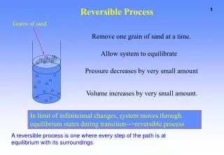
Definition • Severe headaches with or without seizures or neurologic deficits and constriction of cerebral arteries which resolves spontaneously within 1-3 months
Synonyms or Included Disorders • Isolated benign cerebral vasculitis or angiopathy • Call-Fleming syndrome • CNS pseudovasculitis • Benign angiopathy of the central nervous system • Postpartum angiopathy • Migrainous vasospasm • Migraine angiitis • Idiopathic thunderclap headache with reversible vasospasm • Drug induced cerebral vasculopathy • Fatal vasospasm in migrainous infarction
Epidemiology • Females > males (2-10:1) • Mean age of onset = 45 y.o. • Incidence unknown -- probably under diagnozed especially purely cepahalalgic form • Up to 60% are “secondary”
Clinical Presentation • Headache (secondary) - “thunderclap variety”, peaks within one minute and very intense • Only symptom in 75% • Multiple over 1-4 week period is almost pathognomonic • Usually posterior and bilateral • Nausea/vomiting, photophobia, phonophobia • Focal neurologic deficits and seizures in minority of patients
Complications • Localized cortical SAH (20-25%) • Ischemic or hemorrhagic stroke (5-10%) • PRES • Permanent sequelae of a usually benign entity
Pathophysiology • Proposed mechanism: transient disturbance of cerebral arterial vascular tone in segmental and multifocal fashion • Leads to various areas of constriction and/or dilatation • Either idiopathic (primary) or secondary (25-60%)
Secondary Causes • Vasoactive sympathomimetic or serotoninergic substances • Selective serotonic uptake inhibitors, alpha-sympathomimetics (nasal decongestants), some diet pills • Illicit drugs: cannabis, cocaine, ecstasy • Postpartum state • Usually 1st week postpartum after normal delivery • 50-70% associated with vasoconstrictors used to treat postpartum hemorrhage or inhibit lactation • Other causes: hypercalcemia, pheochromocytoma, exercise, and sexual intercourse
Differential Diagnosis • Aneurysmal subarachnoid hemorrhage • Correlates with site and severity of vasospasm • Rare isolated to convexity • Cerebral vasculitis, particularly PACNS (Primary angiitis of the central nervous system) • More insidious, gradually progressive headache • Most have MRI abnormalities: multiple, small infarcts • CSF is markedly abnormal • Preferentially affects small-to-medium arteries whereas RCVS affects medium-to-large arteries
More DDx for Thunderclap Headache • Other intracranial hemorrhages (cerebellar and interventricular) • Cervical and intracranial arterial dissections • Intracranial venous thrombosis • Giant cell arteritis • Pituitary apoplexy • Non-vascular disorders: acute sinusitis, meningitis and CSF hypotension
RCVS and PRES • Overlap: about 10% of cases of RCVS are associated with PRES, regardless of cause • Share similar clinical features • PRES has characteristic findings on MRI • Symmetrical white matter edema in posterior cerebral hemispheres, particularly parieto-occipital regions • Hypo- or iso-intense on DWI and hyperintense on ADC map distinguishes it from stroke in most patients
Imaging • Imaging plays a vital role as the condition is defined in part by the reversibility of the cerebral vasoconstriction • Although rarely used, catheter cerebral angiography is considered the “gold standard”
Non-contrast CT • In uncomplicated RCVS: usually normal • May show cortical SAH or intracebral hemorrhage in complicated cases • Should be followed by lumbar puncture if normal to rule out SAH and inflammatory conditions like infection or cerebral vasculitis
MRI • Usually normal • May show evidence of infarctions, especially in “watershed” zones • May look like PRES • Parenchymal hemorrhages or cortical SAH
Axial FLAIR & DWI (top & middle left) show high signal from right cerebellar infarct. MRA (bottom left) suggests vasculitis. Lateral (center) ICA injection & frontal (right) vertebral artery injection show typical “sausage” beading of RVCS.
Post partum patient shows convexity SAH on FLAIR (left), small bleed on T2* (center) & beading of arteries (right) especially in the right posterior cerebral artery.
CTA/MRA/Angiography • Alternating areas of constriction and dilatation – a.k.a. “beading” -- in several vascular territories • May be seen in large-to-medium-sized arteries of anterior or posterior circulation • Abnormalities may be absent early but show up on repeat imaging, believed to start distallyand move centripetally • NOT specific for RCVS • Resolution within 3 months is most specific for RCVS
Post partum patient shows acute right parietal hematoma on CT (left), SAH on FLAIR (center left), PRES-like cerebellar findings (center right) & beading/thinning of arteries on MRA (right).
Prognosis • Highly dependent on the occurrence of stroke (6-9%) • Otherwise, by definition, most resolve completely without any sequelae
Treatment • Symptomatic (pain, seizures, blood pressure control) • Trigger avoidance (either activity or vasoactive substances) • Observation • Calcium channel blockers • IV magnesium • Short-course of steroids?
Sources Calabrese LH, Dodick DW, Schwedt TJ, et al. Narrative review: reversible cerebral vasoconstriction syndromes. Ann Intern Med 2007; 146: 34–44. Ducros A, Boukobza M, Porcher R, et al. The clinical and radiological spectrum of reversible cerebral vasoconstriction syndrome. A prospective series of 67 patients. Brain 2007; 130: 3091–101. Ducros A, Bousser M. Reversible cerebral vasoconstriction sydnome. Pract Neurol 2009; 9: 256–267. Koopman K, Uyttenboogaart M, Luijckx GJ, et al. Pitfalls in the diagnosis of reversible cerebral vasoconstriction syndrome and primary angiitis of the central nervous system. Eur J Neurol 2007; 14: 1085–7. Moskowitz SI, Calabrese LH, Weil RJ. Benign angiopathy of the central nervous system presenting with intracerebral hemorrhage. Surg Neurol 2007; 67: 522–7. Whyte CA, Calabrese LH. Reversible cerebral vasoconstriction syndrome. Headache: the journal of head and face pain. 2009; 49: 597-598.