Download
1 / 1
10 likes | 80 Views
Southern blot analyses confirm successful deletion of the sigE gene in Mycobacterium tuberculosis. Schematic representation of the sigE DNA region in H37Rv compared to the constructed mutant TB218. Results show expected DNA bands supporting the gene deletion.

E N D
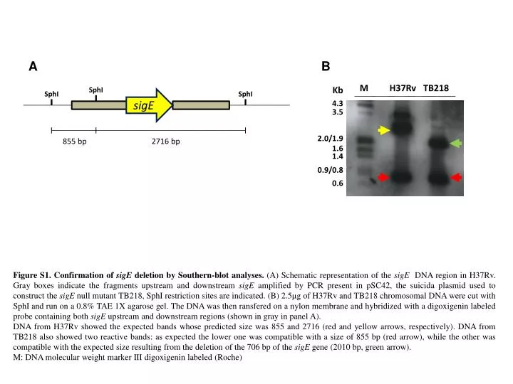
B M H37Rv TB218 Kb SphI SphI SphI A sigE 4.3 3.5 2.0/1.9 855 bp 2716 bp 1.6 1.4 0.9/0.8 0.6 Figure S1. Confirmation of sigEdeletion by Southern-blotanalyses. (A) Schematicrepresentation of the sigEDNA region in H37Rv. Gray boxes indicate the fragmentsupstream and downstream sigEamplified by PCR present in pSC42, the suicida plasmidused to construct the sigEnullmutant TB218, SphIrestrictionsites are indicated. (B) 2.5μg of H37Rv and TB218 chromosomal DNA werecut with SphI and run on a 0.8% TAE 1X agarose gel. The DNA wasthenransfered on a nylon membrane and hybridized with a digoxigeninlabeled probe containingbothsigEupstream and downstream regions (shown in gray in panel A). DNA from H37Rv showed the expectedbandswhosepredictedsizewas 855 and 2716 (red and yellowarrows, respectively). DNA from TB218 alsoshowedtworeactivebands: asexpected the loweronewascompatible with a size of 855 bp (redarrow), while the otherwascompatible with the expectedsizeresulting from the deletion of the 706 bp of the sigEgene (2010 bp, green arrow). M: DNA molecularweight marker III digoxigeninlabeled (Roche)