Download
1 / 18
190 likes | 255 Views
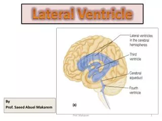
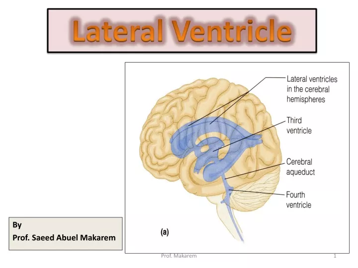
Lateral Ventricle. By Prof. Saeed Abuel Makarem. BRAIN VENTRICLES. The brain is bathed by the cerebrospinal fluid (CSF) Inside the brain, there are spaces (ventricles) filled with CSF There are 4 ventricles 2 lateral ventricles are in the brain hemispheres

E N D
Lateral Ventricle By Prof. Saeed Abuel Makarem Prof. Makarem
BRAIN VENTRICLES • The brain is bathed by the cerebrospinal fluid (CSF) • Inside the brain, there are spaces (ventricles) filled with CSF • There are 4 ventricles • 2 lateral ventriclesare in the brain hemispheres • 3rd ventricle is in the diencephalon • 4th ventricle is between the pons, open medulla and the cerebellum • The 3rd & the 4th ventricles are connected by the cerebral aqueduct Prof. Makarem
Lateral Ventricle • Definition : • It is the cavity of the cerebral hemisphere. • It is C-shaped. • It has 3 horns & central part. • Anterior Horn: in the frontal lobe. • Posterior horn: in the occipital lobe • Inferior horn: in temporal lobe. • Central part or body: in the parietal lobe. Prof. Makarem
Lateral Ventricle • Superior view of the ventricular system. • Lateral ventricle • Anterior horn in the frontal lobe. • Posterior horn in the occipital lobe. • Inferior horn in the temporal lobe. • Body: In the parietal lobe. • The inferior and posterior horns are connected in the trigon. Prof. Makarem
Anterior Horn • In the frontal lobe. • Roof: • Corpus callosum (trunk) • Floor: • Corpus callosum (Rostrum) • Anterior: • Corpus callosum(Genu) • Medially: • Septum pellucidum. • Laterally: • Head of Caudate nucleus. Prof. Makarem
Body or Central part • Lies in the parietal lobe. • Roof: • Corpus callosum (Trunk). • Floor: • Sloping, From lateral to medial it is formed by: • Body of caudate nucleus, • Upper surface of thalamus • Choroid plexus, • Body of fornix. • Medial wall: • Septum pellucidum. • Lateral wall: • narrow area at the meeting of roof & floor. Prof. Makarem
Posterior Horn • In the occipital lobe. • Roof, lateral wall, and floor: • Are formed by the Tapetum of the corpus callosum. • Medially: • There are 2 elevations: Bulb of posterior horn (formed by forceps major-2-). Calcar avis: produced by calcarine sulcus-3-. Prof. Makarem
Inferior Horn • It lies in the temporal lobe. • Roof: • Tapetum, • Tail of caudate nucleus, • Amygdaloid nucleus • Stria terminalis. • Floor: • Hippocampus, • Fimbria of hippocampus & Collateral eminence. • Lateral wall: • Tapetum of the corpus callosum. Prof. Makarem
3rd VENTRICLE Prof. Makarem
Third ventricle is a narrow slit-like cavity whose lateral walls are formed by the thalamus and hypothalamus on either side. At the rostral margin of the midbrain, the cerebral aqueduct opens into the third ventricle. Prof. Makarem
The roof of the ventricle is formed by pia-ependyma, which spans between the two striae medullaris thalami, situated along the dorsomedial border of the thalamus. Prof. Makarem
In the rostral part of the third ventricle lies an aperture, the interventricular foramen or foramen of Monro, which is located between the column of the fornix and the anterior pole of the thalamus. Prof. Makarem
TOPOGRAPHICAL ANATOMY Prof. Makarem
The third ventricle is a midline, slit-like cavity. Prof. Makarem
The lateral walls of the 3rd ventricle is formed of the thalamus and hypothalamus. • Caudally, the third ventricle becomes continuous with thecerebral aqueduct. Prof. Makarem
The interventricular foramenprovides communication, on either side, with the extensive lateral ventricle located within the cerebral hemisphere. Prof. Makarem