Download
1 / 45
450 likes | 470 Views
Learn about the anatomy, functions, and regulation of the pancreas in the digestive system. Discover how pancreatic juice aids in food breakdown, neutralizes chyme, and regulates enzyme release.

E N D
The Digestive System Part C 23
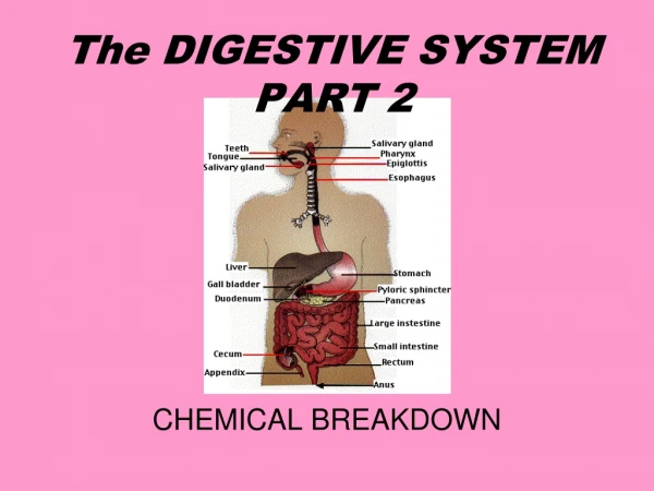
Pancreas • Location • Lies deep to the greater curvature of the stomach • The head is encircled by the duodenum and the tail abuts the spleen
Pancreas • Exocrine function • Secretes pancreatic juice which breaks down all categories of foodstuff • Acini (clusters of secretory cells) contain zymogen granules with digestive enzymes • The pancreas also has an endocrine function – release of insulin and glucagon
Acinus of the Pancreas Figure 23.26a
Composition and Function of Pancreatic Juice • Water solution of enzymes and electrolytes (primarily HCO3–) • Neutralizes acid chyme • Provides optimal environment for pancreatic enzymes • Enzymes are released in inactive form and activated in the duodenum
Composition and Function of Pancreatic Juice • Examples include • Trypsinogen is activated to trypsin • Procarboxypeptidase is activated to carboxypeptidase • Active enzymes secreted • Amylase, lipases, and nucleases • These enzymes require ions or bile for optimal activity
Regulation of Pancreatic Secretion • Secretin and CCK are released when fatty or acidic chyme enters the duodenum • CCK and secretin enter the bloodstream • Upon reaching the pancreas: • CCK induces the secretion of enzyme-rich pancreatic juice • Secretin causes secretion of bicarbonate-rich pancreatic juice • Vagal stimulation also causes release of pancreatic juice
Regulation of Pancreatic Secretion Figure 23.28
Digestion in the Small Intestine • As chyme enters the duodenum: • Carbohydrates and proteins are only partially digested • No fat digestion has taken place
Digestion in the Small Intestine • Digestion continues in the small intestine • Chyme is released slowly into the duodenum • Because it is hypertonic and has low pH, mixing is required for proper digestion • Required substances needed are supplied by the liver • Virtually all nutrient absorption takes place in the small intestine
Motility in the Small Intestine • The most common motion of the small intestine is segmentation • It is initiated by intrinsic pacemaker cells (Cajal cells) • Moves contents steadily toward the ileocecal valve • After nutrients have been absorbed: • Peristalsis begins with each wave starting distal to the previous • Meal remnants, bacteria, mucosal cells, and debris are moved into the large intestine
Control of Motility • Local enteric neurons of the GI tract coordinate intestinal motility • Cholinergic neurons cause: • Contraction and shortening of the circular muscle layer • Shortening of longitudinal muscle • Distension of the intestine
Control of Motility • Other impulses relax the circular muscle • The gastroileal reflex and gastrin: • Relax the ileocecal sphincter • Allow chyme to pass into the large intestine
Large Intestine • Has three unique features: • Teniae coli – three bands of longitudinal smooth muscle in its muscularis • Haustra – pocketlike sacs caused by the tone of the teniae coli • Epiploic appendages – fat-filled pouches of visceral peritoneum
Large Intestine • Is subdivided into the cecum, appendix, colon, rectum, and anal canal • The saclike cecum: • Lies below the ileocecal valve in the right iliac fossa • Contains a wormlike vermiform appendix
Large Intestine Figure 23.29a
Colon • Has distinct regions: ascending colon, hepatic flexure, transverse colon, splenic flexure, descending colon, and sigmoid colon • The transverse and sigmoid portions are anchored via mesenteries called mesocolons • The sigmoid colon joins the rectum • The anal canal, the last segment of the large intestine, opens to the exterior at the anus
Valves and Sphincters of the Rectum and Anus • Three valves of the rectum stop feces from being passed with gas • The anus has two sphincters: • Internal anal sphincter composed of smooth muscle • External anal sphincter composed of skeletal muscle • These sphincters are closed except during defecation
Mesenteries of Digestive Organs Figure 23.30b
Mesenteries of Digestive Organs Figure 23.30c
Mesenteries of Digestive Organs Figure 23.30d
Large Intestine: Microscopic Anatomy • Colon mucosa is simple columnar epithelium except in the anal canal • Has numerous deep crypts lined with goblet cells • Anal canal mucosa is stratified squamous epithelium • Anal sinuses exude mucus and compress feces • Superficial venous plexuses are associated with the anal canal • Inflammation of these veins results in itchy varicosities called hemorrhoids
Structure of the Anal Canal Figure 23.29b
Bacterial Flora • The bacterial flora of the large intestine consist of: • Bacteria surviving the small intestine that enter the cecum and • Those entering via the anus • These bacteria: • Colonize the colon • Ferment indigestible carbohydrates • Release irritating acids and gases (flatus) • Synthesize B complex vitamins and vitamin K
Functions of the Large Intestine • Other than digestion of enteric bacteria, no further digestion takes place • Vitamins, water, and electrolytes are reclaimed • Its major function is propulsion of fecal material toward the anus • Though essential for comfort, the colon is not essential for life
Motility of the Large Intestine • Haustral contractions • Slow segmenting movements that move the contents of the colon • Haustra sequentially contract as they are stimulated by distension • Presence of food in the stomach: • Activates the gastrocolic reflex • Initiates peristalsis that forces contents toward the rectum
Defecation • Distension of rectal walls caused by feces: • Stimulates contraction of the rectal walls • Relaxes the internal anal sphincter • Voluntary signals stimulate relaxation of the external anal sphincter and defecation occurs
Defecation Figure 23.32
Chemical Digestion: Carbohydrates • Absorption: via cotransport with Na+, and facilitated diffusion • Enter the capillary bed in the villi • Transported to the liver via the hepatic portal vein • Enzymes used: salivary amylase, pancreatic amylase, and brush border enzymes
Chemical Digestion: Proteins • Absorption: similar to carbohydrates • Enzymes used: pepsin in the stomach • Enzymes acting in the small intestine • Pancreatic enzymes – trypsin, chymotrypsin, and carboxypeptidase • Brush border enzymes – aminopeptidases, carboxypeptidases, and dipeptidases
Chemical Digestion: Proteins Figure 23.34
Chemical Digestion: Fats • Absorption: Diffusion into intestinal cells where they: • Combine with proteins and extrude chylomicrons • Enter lacteals and are transported to systemic circulation via lymph • Glycerol and short chain fatty acids are: • Absorbed into the capillary blood in villi • Transported via the hepatic portal vein • Enzymes/chemicals used: bile salts and pancreatic lipase
Chemical Digestion: Fats Figure 23.35
Fatty Acid Absorption • Fatty acids and monoglycerides enter intestinal cells via diffusion • They are combined with proteins within the cells • Resulting chylomicrons are extruded • They enter lacteals and are transported to the circulation via lymph
Fatty Acid Absorption Figure 23.36
Chemical Digestion: Nucleic Acids • Absorption: active transport via membrane carriers • Absorbed in villi and transported to liver via hepatic portal vein • Enzymes used: pancreatic ribonucleases and deoxyribonuclease in the small intestines
Electrolyte Absorption • Most ions are actively absorbed along the length of small intestine • Na+ is coupled with absorption of glucose and amino acids • Ionic iron is transported into mucosal cells where it binds to ferritin • Anions passively follow the electrical potential established by Na+
Electrolyte Absorption • K+ diffuses across the intestinal mucosa in response to osmotic gradients • Ca2+ absorption: • Is related to blood levels of ionic calcium • Is regulated by vitamin D and parathyroid hormone (PTH)
Water Absorption • 95% of water is absorbed in the small intestines by osmosis • Water moves in both directions across intestinal mucosa • Net osmosis occurs whenever a concentration gradient is established by active transport of solutes into the mucosal cells • Water uptake is coupled with solute uptake, and as water moves into mucosal cells, substances follow along their concentration gradients
Malabsorption of Nutrients • Results from anything that interferes with delivery of bile or pancreatic juice • Factors that damage the intestinal mucosa (e.g., bacterial infection) • Gluten enteropathy (adult celiac disease) – gluten damages the intestinal villi and reduces the length of microvilli • Treated by eliminating gluten from the diet (all grains but rice and corn)
Embryonic Development of the Digestive System • 3rd week – endoderm has folded and foregut and hindgut have formed • The midgut is open and continuous with the yolk sac • Mouth and anal openings are nearly formed • 8th week – accessory organs are budding from endoderm
Embryonic Development of the Digestive System Figure 23.37
Developmental Aspects • During fetal life, nutrition is via the placenta, but the GI tract is stimulated toward maturity by amniotic fluid swallowed in utero • At birth, feeding is an infant’s most important function and is enhanced by • Rooting reflex (helps infant find the nipple) and sucking reflex (aids in swallowing) • Digestive system has few problems until the onset of old age • During old age the GI tract activity declines, absorption is less efficient, and peristalsis is slowed
Cancer • Stomach and colon cancers rarely have early signs or symptoms • Metastasized colon cancers frequently cause secondary liver cancer • Prevention is by regular dental and medical examinations
Cancer • Colon cancer is the 2nd largest cause of cancer deaths in males (lung cancer is 1st) • Forms from benign mucosal tumors called polyps whose formation increases with age • Regular colon examination should be done for all those over 50