Download
1 / 58
580 likes | 593 Views
"Explore the vital functions and physical characteristics of blood, its role in transportation, protection, and regulation within the body. Learn about the major components, such as plasma and formed elements, and understand the significance of hematocrit and hemoglobin levels. Discover the fascinating world of red and white blood cells along with platelets. Get insights into how blood maintains homeostasis, regulates pH, and plays a crucial role in overall health. Find out why donating blood can make a life-saving difference. Join us in understanding the complexities and wonders of this essential bodily fluid."

E N D
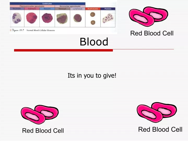
Blood Its in you to give!
Blood • If you took a snapshot of where blood is at any given moment, there would be 10 % is in the lungs, 5% heart, and 85% throughout the rest of the body. • The temperature is 38 degrees C, which is slightly warmer than the overall body temperature.
Physical Characteristics of Blood • Thicker (more viscous) than water and flows more slowly than water • Temperature of 100.4 degrees F • pH 7.4 (7.35-7.45) • 8 % of total body weight • Blood volume • 5 to 6 liters in average male • 4 to 5 liters in average female • hormonal negative feedback systems maintain constant blood volume and osmotic pressure
3 major functions of blood • 1. Transportation • 2. Protection • 3. Regulation
1. Transportation • transporting of all substances necessary for cellular metabolism • This is done through respiration, nutrition and excretion
Transportation - respiration • O2 goes to the tissue via RBCs • O2 is picked up from the caps and binds with Hb (O2 carrying protein in RBC) • O2 is released from the Hb as blood flows through the caps • O2 diffuses into the ISF and then into the cells • CO2 from the tissue cells is released from the Hb and exhaled.
Transportation - Nutritive • Blood has a nutritive function • it carries absorbed products of digestion through the liver to the cells of the body for nourishment
Transportation - excretory • Blood has an excretory function • metabolic wastes, excess water and ions, and other molecules in plasma (fluid aspect of blood) are filtered through blood vessels of the kidneys and excreted in urine
2. Protection • Protection against injury / blood loss (clotting) • Protection against foreign microbes or toxins (infection) introduced into the body • Clotting mechanism protects against blood loss when vessels are damaged • Leukocytes (WBCs) provide immunity - phagocytosis
3. Regulatory - homeostasis • Regulation by carrying hormones • Body Temperature maintenance • pH regulation • Fluid volume regulation, water content in cells
Functions of Blood • Transportation • O2, CO2, metabolic wastes, nutrients, heat & hormones • Regulation • helps regulate pH through buffers • helps regulate body temperature • coolant properties of water • vasodilatation of surface vessels dump heat • helps regulate water content of cells by interactions with dissolved ions and proteins • Protection from disease & loss of blood
Major components of blood • Blood is a type of connective tissue • Cells suspended in the fluid intercellular matrix is called plasma • Cells are RBCs, WBCs, platelets from bone marrow • Blood is thicker than water as you know and you can’t get blood from a stone • Volume varies with body size; ave 70 kg man has 5 L, roughly 10% and varies with changes in the proportions of tissues - more or less fat etc.
Components of Blood • Hematocrit • 55% plasma • 45% cells • 99% RBCs • < 1% WBCs and platelets
1. Plasma 2. Formed elements Major components of blood
Plasma • Functions is to transport nutrients, gases, vitamins, regulate fluid electrolytes and balance pH • Blood plasma is a clear pale yellow fluid • Mostly water dissolved proteins, hormones, nutrients, minerals and wastes
Formed Elements of Blood • Red blood cells ( erythrocytes ) • White blood cells ( leukocytes ) • granular leukocytes • neutrophils • eosinophils • basophils • agranular leukocytes • lymphocytes = T cells, B cells, and natural killer cells • monocytes • Platelets (special cell fragments)
Hematocrit • Percentage of blood occupied by cells • female normal range • 38 - 46% (average of 42%) • male normal range • 40 - 54% (average of 46%) • testosterone • Anemia • not enough RBCs or not enough hemoglobin • Polycythemia • too many RBCs (over 65%) • dehydration, tissue hypoxia, blood doping in athletes
Hemoglobin • Globin protein consisting of 4 polypeptide chains • One heme pigment attached to each polypeptide chain • each heme contains an iron ion (Fe+2) that can combine reversibly with one oxygen molecule
Red Blood Cells or Erythrocytes • Contain oxygen-carrying protein hemoglobin that gives blood its red color • 1/3 of cell’s weight is hemoglobin • Biconcave disk 8 microns in diameter • increased surface area/volume ratio • flexible shape for narrow passages • no nucleus or other organelles • no cell division or mitochondrial ATP formation • Normal RBC count • male 5.4 million/drop ---- female 4.8 million/drop • new RBCs enter circulation at 2 million/second
Because hemoglobin is a major component and this reflects the capability of blood to carry O2, it is often measured medically. • Polycythemia is an increased Hb level • Anemia is a decreased Hb level • When you want to know the % of RBCs in the blood is known as hematocrit (HCT) • RBC count - 5.4 million/mm3 for a male 4.8 million/mm3 female
Transport of O2, CO2 and Nitric Oxide • Each hemoglobin molecule can carry 4 oxygen molecules from lungs to tissue cells • Hemoglobin transports 23% of total CO2 waste from tissue cells to lungs for release • combines with amino acids in globin portion of Hb • Hemoglobin transports nitric oxide & super nitric oxide helping to regulate BP • iron ions pick up nitric oxide (NO) & super nitric oxide (SNO)& transport it to & from the lungs • NO causing vasoconstriction is released in the lungs • SNO causing vasodilation is picked up in the lungs
RBC Life Cycle • RBCs live only 120 days • wear out from bending to fit through capillaries • no repair possible due to lack of organelles • Worn out cells removed by fixed macrophages in spleen & liver • Breakdown products are recycled • Bilrubin is one of the breakdown products and is a yellow pigment
Leukocytes or WBCs • Main role is phagocytosis and antibody production • WBCs are granular or agranular based upon the ability of the cell or hold gram stain in the lab
WBC Anatomy and Types • All WBCs (leukocytes) have a nucleus and no hemoglobin • Granular or agranular classification based on presence of cytoplasmic granules made visible by staining • granulocytes are neutrophils, eosinophils or basophils • agranulocytes are monocyes or lymphocytes
Leukocytes or WBCs • Granular WBCs - red/orange cytoplasm granules • The shape of the nucleus and staining properties of the cytoplasm granules distinguish one WBC from the other • There are 3 types of granular WBCs • 1. Eosinophils • 2. Basophils • 3. Neutrophils
Eosinophils -allergic reactions - immune complexes curing allergic reaction lessening the severity of the reaction, inactivates chemicals released with allergic reactions, phagocytosis, parasite elimination WBCs
Eosinophils (Granulocyte) • Nucleus with 2 or 3 lobes connected by a thin strand • Large, uniform-sized granules stain orange-red with acidic dyes • do not obscure the nucleus • Diameter is 10 to 12 microns • 2 to 4% of circulating WBCs
Eosinophil Function • Leave capillaries to enter tissue fluid • Release histaminase • slows down inflammation caused by basophils • Attack parasitic worms • Phagocytize antibody-antigen complexes
Basophils - release chemicals (heparin for anticoagulation and histamine for inflammatory control) during allergic reactions WBCs
Basophils (Granulocyte) • Large, dark purple, variable-sized granules stain with basic dyes • obscure the nucleus • Irregular, s-shaped, bilobed nuclei • Diameter is 8 to 10 microns • Less than 1% of circulating WBCs
Basophil Function • Involved in inflammatory and allergy reactions • Leave capillaries & enter connective tissue as mast cells • Release heparin, histamine & serotonin • heighten the inflammatory response and account for hypersensitivity (allergic) reaction
Neutrophils (Granulocyte) • Polymorphonuclear Leukocytes or Polys (PMN’s) • Nuclei = 2 to 5 lobes connected by thin strands • older cells have more lobes • young cells called band cells because of horseshoe shaped nucleus (band) • Fine, pale lilac practically invisible granules • Diameter is 10-12 microns • 60 to 70% of circulating WBCs
Neutrophil Function • Fastest response of all WBC to bacteria • Direct actions against bacteria • release lysozymes which destroy/digest bacteria • release defensin proteins that act like antibiotics & poke holes in bacterial cell walls destroying them • release strong oxidants (bleach-like, strong chemicals ) that destroy bacteria
Agranular WBCs • round / kidney shaped nucleus • possess cytoplasm granules but are not visible with the light microscope because of small or poor staining quality • 2 types – Lymphocytes & Monocytes
Lymphocytes - natural killer cells Monocytes - macrophages during phagocytosis Agranular WBCs
Lymphocyte (Agranulocyte) • Dark, oval to round nucleus • Cytoplasm sky blue in color • amount varies from rim of blue to normal amount • Small cells 6 - 9 microns in diameter • Large cells 10 - 14 microns in diameter • increase in number during viral infections • 20 to 25% of circulating WBCs
Lymphocyte Functions • B cells • destroy bacteria and their toxins • turn into plasma cells that produces antibodies • T cells • attack viruses, fungi, transplanted organs, cancer cells & some bacteria • Natural killer cells • attack many different microbes & some tumor cells • destroy foreign invaders by direct attack
Monocyte (Agranulocyte) • Nucleus is kidney or horse-shoe shaped • Largest WBC in circulating blood • does not remain in blood long before migrating to the tissues • differentiate into macrophages • fixed group found in specific tissues • alveolar macrophages in lungs • kupffer cells in liver • wandering group gathers at sites of infection • Diameter is 12 - 20 microns • Cytoplasm is a foamy blue-gray • 3 to 8% o circulating WBCs
Monocyte Function • Take longer to get to site of infection, but arrive in larger numbers • Become wandering macrophages, once they leave the capillaries • Destroy microbes and clean up dead tissue following an infection
Differential WBC Count • Detection of changes in numbers of circulating WBCs (percentages of each type) indicates infection, poisoning, leukemia, chemotherapy, parasites or allergy reaction • Normal WBC counts • neutrophils 60-70% (up if bacterial infection) • lymphocyte 20-25% (up if viral infection) • monocytes 3 -- 8 % (up if fungal/viral infection) • eosinophil 2 -- 4 % (up if parasite or allergy reaction) • basophil <1% (up if allergy reaction or hypothyroid)
Complete Blood Count • Screens for anemia and infection • Total RBC, WBC & platelet counts; differential WBC; hematocrit and hemoglobin measurements • Normal hemoglobin range • infants have 14 to 20 g/100mL of blood • adult females have 12 to 16 g/100mL of blood • adult males have 13.5 to 18g/100mL of blood
Platelet (Thrombocyte) Anatomy • Disc-shaped, 2 - 4 micron cell fragment with no nucleus • Normal platelet count is 150,000-400,000/drop of blood • Other blood cell counts • 5 million red & 5-10,000 white blood cells
Hemostasis • Stoppage of bleeding in a quick & localized fashion when blood vessels are damaged • Prevents hemorrhage (loss of a large amount of blood) • Methods utilized 3 • vascular spasm • platelet plug formation • blood clotting (coagulation = formation of fibrin threads)
Vascular Spasm • Damage to blood vessel produces stimulates pain receptors • Reflex contraction of smooth muscle of small blood vessels • Can reduce blood loss for several hours until other mechanisms can take over • Only for small blood vessel or arteriole
Platelet Plug Formation • Platelets store a lot of chemicals in granules needed for platelet plug formation • alpha granules • clotting factors • platelet-derived growth factor • cause proliferation of vascular endothelial cells, smooth muscle & fibroblasts to repair damaged vessels • dense granules • ADP, ATP, Ca+2, serotonin, fibrin-stabilizing factor, & enzymes that produce thromboxane A2 • Steps in the process • (1) platelet adhesion (2) platelet release reaction (3) platelet aggregation
Hemostasis • the process of stopping the flow of blood from a broken or damaged blood vessel • 1. Constriction of the BV • 2. Formation of platelet plug • 3. Coagulation
1. Constriction of the BV • When BV is severed or damaged contraction of smooth ms in vessel wall contracts the vessel at its damaged end decreasing the size of the opening blood can escape through • Vasoconstriction or vasospasm • Lasts up to 30 mins when by then the other processes are in play