Download

1 / 70
700 likes | 716 Views
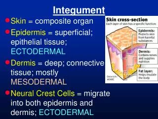
INTEGUMENT-1. This resource is licensed under the Creative Commons Attribution Non-Commercial & No Derivative Works License. Objectives. To identify the main features of non-hairy heavily keratinised skin and to compare with thinner hairy skin. 2. To identify sebaceous and sweat glands.

E N D
INTEGUMENT-1 This resource is licensed under the Creative Commons Attribution Non-Commercial & No Derivative Works License
Objectives To identify the main features of non-hairy heavily keratinised skin and to compare with thinner hairy skin. 2. To identify sebaceous and sweat glands.
SLIDE 77 Digital skin (monkey) Examine this section under very low magnification, (Masson’s trichrome stain). Skin is a difficult structure to cut for histology; the outer keratinised layers often pull apart or tear during sectioning. Part of the epidermis in the section below has been lost (arrowed). 1.0 mm
SLIDE 77 Digital skin (monkey) Identify - thick stratum corneum. What colour does it stain? - rest of the epidermis. What colour does it stain? - dermis - hypodermis 250 µm
SLIDE 77 Digital skin (monkey) hypodermis dermis Identify - thick stratum corneum. What colour does it stain? Red. - rest of the epidermis. What colour does it stain? Purple. - dermis - hypodermis stratum corneum other epidermal layers 250 µm
SLIDE 77 Digital skin (monkey) What glands and their ducts can be seen in this section? 250 µm
SLIDE 77 Digital skin (monkey) S : sweat glands S S What glands and their ducts can be seen in this section? Sweat glands. What is the predominant tissue in the hypodermis? ducts from sweat glands S 250 µm
SLIDE 77 Digital skin (monkey) S : sweat glands A : adipose tissue in hypodermis S A S What glands and their ducts can be seen in this section? Sweat glands. What is the predominant tissue in the hypodermis? Adipose tissue. ducts from sweat glands S 250 µm
SLIDE 77 Digital skin (monkey) The stain used is Masson’s Trichrome. Dermal papillae interdigitating with the epidermal layers, strengthen the layers. What is stained blue-green in the dermis? 100 µm
SLIDE 77 Digital skin (monkey) D D : dermal papillae D D The stain used is Masson’s Trichrome. Dermal papillae interdigitating with the epidermal layers, strengthen the layers. What is stained blue-green in the dermis? The collagen fibres in the dermis stain blue-green. D epidermis D D dermis 100 µm sweat gland
SLIDE 77 Digital skin (monkey) Label the five strata in the epidermis. 50 µm
SLIDE 77 Digital skin (monkey) stratum corneum stratum lucidum Label the five strata in the epidermis. stratum granulosum stratum spinosum stratum basale 50 µm
Label the five strata in the epidermis. In which of these layers might one observe (a) mitosis? (b) melanocyte (cell bodies)? SLIDE 77 Digital skin (monkey) stratum corneum stratum lucidum Label the five strata in the epidermis. stratum granulosum stratum spinosum stratum basale 50 µm
Label the five strata in the epidermis. In which of these layers might one observe (a) mitosis? Stratum basale. (b) melanocyte (cell bodies)? SLIDE 77 Digital skin (monkey) stratum corneum stratum lucidum Label the five strata in the epidermis. stratum granulosum stratum spinosum stratum basale (The stratum lucidum is often difficult to distinguish). 50 µm
Label the five strata in the epidermis. In which of these layers might one observe (a) mitosis? Stratum basale. (b) melanocyte (cell bodies)? Stratum basale. SLIDE 77 Digital skin (monkey) stratum corneum stratum lucidum Label the five strata in the epidermis. stratum granulosum stratum spinosum stratum basale (The stratum lucidum is often difficult to distinguish). 50 µm
Sensory Receptors in Skin(revision from nerve cell histology class)
SLIDE 77 Skin to observe Sensory Receptorsdigital skin of monkey Most of the sensory nerve fibres in the skin have free, uncapsulated endings and are concerned with detecting temperature, pain etc . 1.0 mm
SLIDE 77 Skin to observe Sensory Receptorsdigital skin of monkey Pacinian corpuscles are found in the hypodermis. 1.0 mm
SLIDE 77 Skin to observe Sensory Receptors Pacinian corpuscle. 250 µm
SLIDE 77 Skin to observe Sensory Receptors Pacinian corpuscle. Pacinian corpuscle hypodermis dermis epidermis 250 µm
SLIDE 77 Skin to observe Sensory Receptors Pacinian corpuscle. 50 µm
SLIDE 77 Skin to observe Sensory Receptorsdigital skin of monkey Meissner’s corpuscles are found in the dermal papillae. 1.0 mm
SLIDE 77 Skin to observe Sensory Receptors Meissner’s corpuscle. 25 µm
SLIDE 76 Footpad (dog) 1.0 mm Another example of thick skin. This section has been stained with H&E. The keratinised stratum corneum forms a thick outer layer.
SLIDE 76 Footpad (dog) dermal papillae epidermis Another example of thick skin. This section has been stained with H&E. The keratinised stratum corneum forms a thick outer layer. dermis sweat glands hypodermis 1.0 mm
SLIDE 76 Footpad (dog) Higher magnification showing the very thick stratum corneum. Note the spiralling ducts from sweat glands passing through this layer. 250 µm
SLIDE 76 Footpad (dog) stratum corneum Higher magnification showing the very thick stratum corneum. Note the spiralling ducts from sweat glands passing through this layer. ducts from sweat glands 250 µm
SLIDE 76 Footpad (dog) Again identify the five strata in the epidermis. 50 µm
Stratum : SLIDE 76 Footpad (dog) 1 : basale Again identify the five strata in the epidermis. 1 50 µm
Stratum : SLIDE 76 Footpad (dog) 1 : basale 2 : spinosum Again identify the five strata in the epidermis. 1 2 50 µm
Stratum : SLIDE 76 Footpad (dog) 1 : basale 2 : spinosum 3 : granulosum Again identify the five strata in the epidermis. 1 3 2 50 µm
Stratum : SLIDE 76 Footpad (dog) 1 : basale 2 : spinosum 3 : granulosum Again identify the five strata in the epidermis. 4 : lucidum 1 3 4 2 50 µm
Stratum : SLIDE 76 Footpad (dog) 1 : basale 5 2 : spinosum 3 : granulosum Again identify the five strata in the epidermis. 4 : lucidum 1 3 4 5 : corneum 2 50 µm
Keratinised layer of thick skin – electron micrograph Keratinised squamous cells of the stratum corneum : note the lack of nuclei and other cell organelles. The stratum granulosum contains keratohyalin granules and keratin filaments. • Stratum corneum • Stratum granulosum 1 2
The epidermis of the dog is at its thinnest on the ventral side of the abdomen. Hair follicles are visible, cut at varying angles from TS to LS. SLIDE 83 Ventral abdominal skin (dog) 1.0 mm
The epidermis of the dog is at its thinnest on the ventral side of the abdomen. Hair follicles are visible, cut at varying angles from TS to LS. SLIDE 83 Ventral abdominal skin (dog) epidermis H H H : hair follicles dermis hypodermis 1.0 mm
oblique section through hair follicle SLIDE 83 Ventral abdominal skin (dog) epidermal layers transverse section through hair follicle dermis 250 µm
Glands associated with the hair follicle SLIDE 83 Ventral abdominal skin (dog) 100 µm
Glands associated with the hair follicle SLIDE 83 Ventral abdominal skin (dog) sebaceous glands hair follicle duct from apocrine sweat gland 100 µm
Surface of thin skin. Note although the surface is not flat, there are no dermal papillae. The stratum corneum and stratum spinosum are clear, but only a few cells deep. Stratum granulosum not prominent. SLIDE 83 Ventral abdominal skin (dog) 50 µm
Surface of thin skin. Note although the surface is not flat, there are no dermal papillae. The stratum corneum and stratum spinosum are clear, but only a few cells deep. Stratum granulosum not prominent. SLIDE 83 Ventral abdominal skin (dog) stratum granulosum stratum spinosum stratum corneum stratum basale 50 µm
Apocrine sweat glands close to hair follicle. SLIDE 83 Ventral abdominal skin (dog) open lumen of sweat gland lined by a low epithelium 25 µm
SLIDE 74 Dorsal skin (dog) Hairs are more numerous on the dorsal skin of the dog. Compound hair follicles (as in the ventral skin). 1.0 mm
SLIDE 74 Dorsal skin (dog) epidermis dermis hair follicles Hairs are more numerous on the dorsal skin of the dog. Compound hair follicles (as in the ventral skin). hypodermis muscle (in deep layers) 1.0 mm
What is the arrector pili muscle? SLIDE 74 Dorsal skin (dog) 250 µm
What is the arrector pili muscle? A smooth muscle running from below the epidermis and attaching to the root sheath of the hair. SLIDE 74 Dorsal skin (dog) R : root sheath epidermis arrector pili muscle R 250 µm
Compound hair follicles in the dorsal skin of the dog. SLIDE 74 Dorsal skin (dog) 250 µm
Compound hair follicles in the dorsal skin of the dog. SLIDE 74 Dorsal skin (dog) thin epidermis compound hair follicles 250 µm
SLIDE 74 Dorsal skin (dog) The epidermis from the dorsal region skin. Again no evidence of dermal papillae and very few layers of cells present. 50 µm
SLIDE 74 Dorsal skin (dog) stratum corneum The epidermis from the dorsal region skin. Again no evidence of dermal papillae and very few layers of cells present. other epidermal layers dermis 50 µm