Download
1 / 63
630 likes | 936 Views
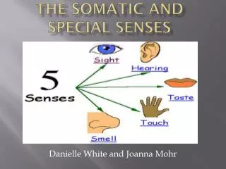
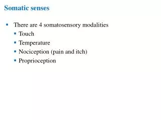
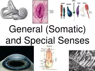
Somatic and Special Senses. Special Senses . General senses. Smell Taste Vision Hearing Balance . Tactile Touch Pressure Thermal (hot vs cold) Pain Proprioceptive. Sensory receptors. Detect environmental changes and trigger nerve impulse Neurons have specific job E.x.

E N D
Special Senses General senses • Smell • Taste • Vision • Hearing • Balance • Tactile • Touch • Pressure • Thermal (hot vs cold) • Pain • Proprioceptive
Sensory receptors • Detect environmental changes and trigger nerve impulse • Neurons have specific job • E.x
Adaptation • Sensors adapt by • Decrease in responsiveness • Perception of stimulus fades/disappears • Fast adapting • Signal change • E.x • Slow adapting • Trigger impulses if stimulus persists • E.x.
Types of Receptors • Mechanoreceptors • Touch, pressure, hearing • Nociceptors • pain • Photoreceptors • light • Chemoreceptors • Chemicals in nose and mouth • Osmoreceptors • Osmotic pressure
Types of mechanoreceptors • Merkel receptors • Sense fine detail • Fires continuously • Meissner corpuscle • Control hand grip • Fire when stimulus is added and removed
Types of mechanoreceptors(Continued) • Ruffini corpuscle • Sensitive to stretching skin • Fire continuously • Pacinian corpuscle • Respond to fine detail when moving fingers • Fire when stimulus is applied and removed
Why is a small portion of your cerebral cortex devoted to your arm while a large portion is devoted to your hands and fingers? • The hands and fingers are more useful for gathering information • Able to feel fine details with fingers Homunucleus Analysis
Pressure vs touch • Pressure is a sustained sensation • Large area • Deeper tissues • Touch receptors are stimulated • Limited area
Other sensations • Itch & tickling • Both arise from stimulation of free nerve endings • Hot & cold • Cold receptors (epidermis) • Hot receptors (dermis) • Both work in specific temp range • What happens when they are exposed to extreme temps?
pain • Fast Pain • Localized • E.x • Slow Pain • Localized in large area • E.x • Why can’t the brain feel pain? • Because is does not contain nocicptors • http://www.youtube.com/watch?v=tnABHy6tjL8
Homunucleus • Is a map that corresponds body part to touch sensitivity • Is this proportional? • Would you expect everyone to have the same image?
Why isn’t your skin’s sensitivity in proportion to the size of the body part? Is it reversed? • How different is your homunucleusfrom your partners? Where are they different and where are they the same? • What would happen if your skin’s sensitivity in your hands stopped working? Make your own homonucleus Analysis Questions
Does cancer have an odor? Dogs • http://www.youtube.com/watch?v=H7mI5Jj9aAQ • http://sciencenetlinks.com/science-news/science-updates/cancer-sniffing-dogs/ Training • http://www.youtube.com/watch?v=IZA9R0uSGWc
olfaction(AKA Sense of smell) • Which structure detects an odor? • Olfactory receptor Olfactory Epithelium • Olfactory receptor • Have hair like extensions • Basal cells • Stem cells that go through cell division
Olfactory Pathway • https://www.youtube.com/watch?v=snJnO6OpjCs • How do we detect an odor? • Odor • Receptor • Olfactory bulb • Olfactory tract • Limbic system or temporal lobe
Gustation(AkA Sense of taste) • Sour • Sweet • Bitter • Salty • Umami (savory/meaty) • How does a cold affect your sense of taste?
Taste Buds(Papillae) • Cover the surface of the tongue • Most are found on the tongue Gustatory Pathway • Taste receptors • Cranial nerves • Medulla oblongata • Limbic system • Parietal lobe
How the brain understands taste • Caused by the release of a neurotransmitter • Why do foods taste different? • By activating different groups of neurons
Mapping taste buds • Where are the receptors for salt found? • Where are the receptors for sweet found? • Where are the receptors for sour found? • Where are the receptors for bitter found? • Where are the receptors for savory found?
Vision • Accessory structures • Eyebrows • Eyelashes • Eyelids • Muscles • Lacrimal apparatus(tears)
Science of tears • What are tears? • Salt, mucus, lysozyme (kills bacteria) • Clean, lubricate and moisten • When do we produce tears? • Parasympathetic stimulation (emotional) • Clear irritants
Layers of the EYEball • Fibrous tunic • Cornea • Sclera • Vascular tunic • Choroid • Ciliary body • iris • Retina
fibrous Tunic • Cornea • Is curved • This varies in individuals and as you age • Sclera • “White” of the eye • Gives shape • Protects • Point of muscle attachment
Vascular Tunic • Choroid • Lines the sclera • Nourishes the retina • Cilliary body • Muscle- controls the shape of the lens • Process- secretes aqueous humor • Aqueous humor • Nourishes the eye as it ciculates through both chambers
Vascular Tunic • Lens • Changes shape to focus light on retina • Clearer vision • Held in place by zonular fibers • Iris • Is convex (curves outward) • Colored part of eye • Pupil • Light enters here • Diameter changes in response to light
The iris is a muscle that controls the size of the pupil. The iris is the colored part of the eye. • In bright light, the iris expands and the pupil gets smaller • In low light, the iris contracts and the pupil gets bigger
Retina • Photoreceptors • Light sensitive cells • Transmit info to brain • E.x. rods and cones • Rods • Low light • Sense shades of grey • Cones • Need brighter light • Sense color
Retina(continued) • Fovea centralis • Sharp central vision • Lots of cones and zero rods • Optic disk • “blind spot” • Does not have photoreceptors (rods and cones)
Inside the eyeball • Vitreous body • Fluid that prevents the eye from collapsing • Intraocular pressure • Refers to fluid inside the eye • Balance between production and drainage of aqueous humor
Muscles of the eyeball • Ciliary muscle • Controls diameter of pupil • Lateral rectus muscle • Moves eye inward • Medial rectus muscle • Moves eye outward • Orbicularis oculi • Open and close the eyelids
. Types of cones • Red cones • Sense red light • Green cones • Sense green light • Blue cones • Sense blue light
Image formation • Refraction • Bending of light rays • Occurs at the cornea and lens • Images on retina • Are inverted • Undergo left-right reversal
Viewing objects • Distant objects (20 ft away) • Light rays are parallel • Curvature is aligned (lens is flat) • Near objects • Light rays are divergent • Lens must increase curvature (rounder) • This is called accommodation
What does 20/20 vision really mean? • At 20 ft away from an object you see what the average person sees
What about 20/40 vision? • When standing 20 ft away from an object you see what someone with normal vision can see when they stand 40 ft away • You need to be closer to the object
What about 20/10 vision? • This means that when you stand 20 feet away from an object you see what the average person sees when they stand 10 feet away from the object • Hawk’s have 20/2 vision, what does this mean?
The retina Two layers • Neural layer • Different types of neurons • E.x. photorecpetors • Pigmented layer • Contains melanin which helps absorb light rays
The visual pathway Images (light rays): • Enter the pupil • Lens inverts imageand projects onto retina • Optic nerve carries message to brain (crosses over at optic chiasm) • Brain interprets image
Binocular Vision • The ability to focus on a set of objects • Depth • 3D vision • Convergence • Occurs when you move forward • Movement of eye inward
Have your partner hold two different pencils at different distances in front of you so that both pencils can be seen. With both eyes open, try to grab the pencil that is furthest from you. Repeat steps one and two a twice. Have your partner change the pencils distance with each trial Repeat steps one through three with one eye closed Testing binocular vision
Structure of the Ear Made up of • External ear • Collects sound waves and moves them inward • Middle ear • Conveys sound to oval window • Internal ear • Receptors for hearing and equilibrium