Download
1 / 31
360 likes | 723 Views
SIALOGRAPHY & THE SALIVARY GLANDS. A radiographic examination of the salivary glands and ducts using contrast media. Injection of contrast media into salivary ducts. OIL BASED CONTRAST SINOGRAPHIN WATER BASED (IONIC). INDICATIONS FOR EXAM. Stones (Calculi) sialolithiasis

E N D
SIALOGRAPHY & THE SALIVARY GLANDS A radiographic examination of the salivary glands and ducts using contrast media
Injection of contrast media into salivary ducts OIL BASED CONTRAST SINOGRAPHIN WATER BASED (IONIC)
INDICATIONS FOR EXAM • Stones (Calculi) sialolithiasis • Obstruction / Strictures • Pain & Swelling • Infection • Masses / Tumors
CONTRAINDICATIONS: • History of contrast media allergies • Parotits (mumps) • Severe inflammation of the salivary ducts
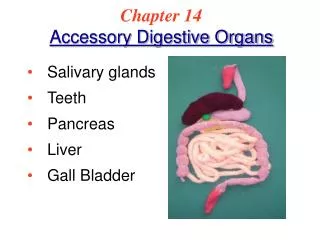
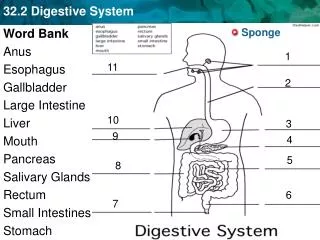
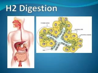
SIALOGRAPHY SNOPEK – CH 21 & BALLINGER – VOL. 2 CH. 14 3 SALIVARY GLANDS – PAROTID – LARGEST – LOCATED BY EAM - MANDIBULAR RAMUS SUBMANDIBULAR / SUBMAXILLARY – 1st molar to Gonion SUBLINGUAL – LOCATED BEHIND THE MENTUM under the sublingual fold SMALLEST-
DUCTS Parotid duct – located by maxillary bone– 2nd upper molar) Stensen’s duct PAROTID GLAND Submandibular Duct – located on end of sublingual ridge to fill submandibular Wharton’s duct SUBMAXILLARY Sublingual duct (several small) located alongside submandibular duct to SUBLINGUAL GLAND
EQUIPMENT NEEDS • RAD/ • FLUORO ROOM Safe and simple but difficult to perform – hard to located and catheterize ducts Mostly replaced now by MRI & CT
PROCEDURE Sialogram Tray – • Gauze, Sterile towels, Spot Light, Magnifying glasses, Gloves, Eyewear, mask • LEMONS - cut into wedges • Lacrimal Probes • Hemostats • 5 cc syringes • Sialogram Catheter (Rabinov) needles or canulas
PATIENT PREP (No specific prep = oral mouthwash in nice) • Remove any metal – bridgework, tongue piercing • Get History – present to radiologist BEFORE setting up tray
32 gauge • Sialogram needle • “Rabinov” catheter
RADIOGRAPHS • 1. SCOUT FILMS – important to r/o stone for SOFT TISSUE (LIGHT EXPOSURE) Like mandible series = AP (OML) (Grid ) • Both Obliques (Ext. cassette) • True Lateral (x-table) (Grid or extremity cassette) • Tangential films may be required • Mentum or Parotid • 2. Give lemon - contast injected • 3. SPOT FILMS – taken by DR during fluoro • 4. Post films – taken 10 min after injections – check drainage
Water based iodinated (ionic or nonionic) Less dense – absorbed faster –no residue Conray, Hypaque, Isovue, Renographin (60/76) Oil-based More dense, absorb slower – can cause granuloma (stones) hard to completely excrete Ethiodol, Sinographin Contrast media
DUCTS • Parotid duct – located by maxillary bone– 2nd upper molar) - PAROTID GLAND Stensen’s duct • Submandibular Duct – located on end of sublingual ridge to fill submandibular / SUBMAXILLARY Wharton’s duct • Sublingual duct (several small) located alongside submandibular duct to SUBLINGUAL GLAND
Tangential parotid gland, supine position. CR - “skims” – side of face Cr is directed along lateral side for unobstructed image of parotid gland
“True” Lateral • Parotid gland is superimposed • Over the mandibular rami
THE END Questions?