Download
1 / 21
220 likes | 518 Views
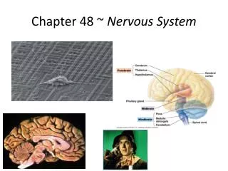
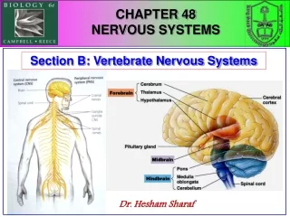
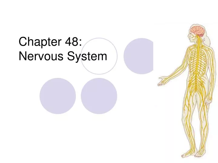
Chapter 48: Nervous System. Neurons and Supporting cells. Organization of Nervous system Diffuse nerve net-hydra Brain in front Central nervous system (CNS)- planarian Ganglia (cluster of neurons)- in annelids Peripheral nervous system (PNS)-connects CNS with rest of body.

E N D
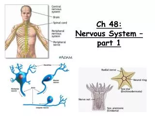
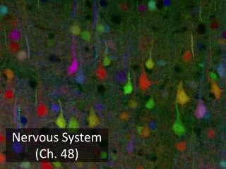
Neurons and Supporting cells • Organization of Nervous system • Diffuse nerve net-hydra • Brain in front • Central nervous system (CNS)- planarian • Ganglia (cluster of neurons)- in annelids • Peripheral nervous system (PNS)-connects CNS with rest of body
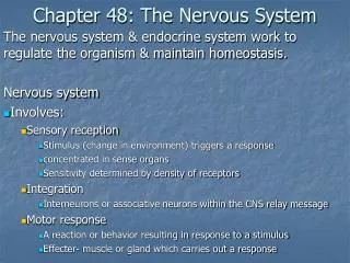
Information Processing • Sensory input- sensory neurons transmit external stimuli or internal conditions to CNS • Integration- interneurons integrate (analyze and interpret). Greatest complexity in neural circuits cells. • Motor output- leaves CNS via motor neurons that communicate with effector cells (muscle or endocrine cells)
Question 1 • a. Arrange the following neurons in the correct sequence for information flow during the knee–jerk reflex: interneuron, sensory neuron, motor neuron. • b. Which of the neuron types is located entirely within the CNS?
Neuron Structure cell body sensory neuron cell body axon interneuron “associative” dendrites dendrites cell body motor neuron
Neuron Structure • Dendrites- receive signals • Synapses- site of communication from presynaptic cell to postsynaptic cell by neurotransmitters • Axon- transmits the signal
Question 2 • Would severing a neuron′s axon stop the neuron from receiving or from transmitting information? Explain.
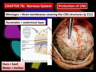
Supporting cells • Astrocytes- structural support • Radial glia- form tracks • Oligodendrocytes (in CNS) and Schwann cells (in PNS) form myelin sheaths around axon • Similar to plastic protecting electric covers
Question 3 • What would be the most obvious structural abnormality in the nervous system of a mouse lacking oligodendrocytes?
Action potentials are the signals that conducted by axons • If gated channels, membrane potential will change when open or closed • Hyperpolarization- increase in membrane potential (inside is more neg) • Ex. Opening of K+ channels • Depolarization- decrease in membrane potential (inside is less neg) • Ex. Opening of Na+ channels
Action potentials cont’d • Threshold- depolarization up to a certain membrane voltage • Action potential-once reached amount of stimulus has been reached; signal that carry information along axons; very brief Animation Link
Question 4 • Draw the sketch below and label the following: outside cell, inside cell. Show where the concentrations of Na+ and K+ are highest.
Conduction of Action Potentials • For an action potential to function long distances, it regenerates itself as it moves along the axon, increasing action potential as it moves down axons • What effects speed of transmission? • Diameter of axon • Myelin sheath
Question 5 • Arrange the following from lowest to highest conduction speed: (a) myelinated, small–diameter axon; (b) myelinated, large–diameter axon; (c) unmyelinated, small–diameter axon.
Neurons communicate with other cells at synapses • Two types of communication: • Electrical synapses- gap junctions that allow electric current to flow from one cell to another • Chemical synapses-most; release neurotransmitter in a synaptic vesicle, it depolarizes the terminal membrane and vesicles fuse with terminal membrane, releasing neurotransmitters by exocytosis. It can then have a direct or indirect effect on postsynaptic cell. Online Link Mouse Party Link
Direct synaptic transmission • Neurotransmitter binds to channel (the receptor), it opens and allows ions to diffuse across the membrane • Result- postsynaptic potential (change in membrane potential) • Excitatory postsynaptic potentials (EPSPs) membrane potential brought down to threshold • Inhibitiory postsynaptic potentials (IPSPs) increase past threshold
Indirect transmission • Neurotransmitter binds to a receptor that is not part of the channel, activates a signal transduction pathway • Slower onset but longer lasting than direct transmission Indirect Link
Types of neurotransmitters • Acetylcholine (Ach)- released at neuromuscular junctions, exciting motor cells; other times, can be an inhibitor • Biogenic amines- neurotransmitters from aa • One group, catecholamines from aa tyrosine include epinephrine and nonepinephrine (hormones); dopamine (brain-affect sleep, mood, attention and learning)
Types of neurotransmitters • Amino acids and peptides • Aa-most in brain • Peptides- short chains called neuropeptides • Gases- some neurons release gases especially NO (nitric oxide) and CO (carbon monoxide) • Aa-most in brain • Peptides- short chains called neuropeptides
Question 6 • A single postsynaptic neuron can be affected by neurotransmitter molecules released by many other neurons, some releasing excitatory and some releasing inhibitory neurotransmitters. What will determine whether an action potential is generated in the postsynaptic neuron?
Question 7 • There are many different types of neurotransmitters. Each neuron secretes only one type of neurotransmitter. Some neurotransmitters hyperpolarize the postsynaptic membrane. Are these excitatory or inhibitory neurotransmitters?